4OPJ

Bh-RNaseH:tcdA-DNA complex


Experimental Data Snapshot

  • Method: X-RAY DIFFRACTION
  • Resolution: 1.54 Å
  • R-Value Free: 0.220 
  • R-Value Work: 0.162 
  • R-Value Observed: 0.167 

wwPDB Validation   3D Report Full Report


This is version 1.2 of the entry. See complete history


Literature

Generating Crystallographic Models of DNA Dodecamers from Structures of RNase H:DNA Complexes.

Egli, M.Pallan, P.S.

(null) Methods Mol Biol 1320: 111-126

  • DOI: https://doi.org/10.1007/978-1-4939-2763-0_8
  • Primary Citation of Related Structures:  
    4OPJ, 4OPK

  • PubMed Abstract: 

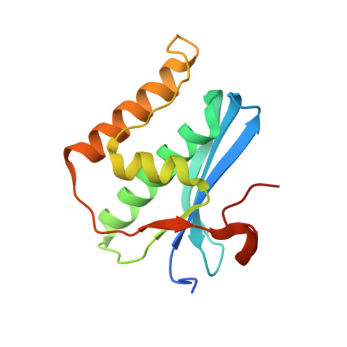
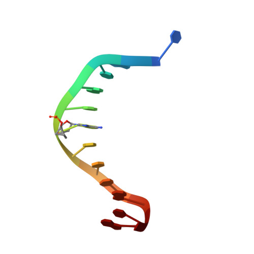
    The DNA dodecamer 5'-d(CGCGAATTCGCG)-3' is arguably the best studied oligonucleotide and crystal structures of duplexes with this sequence account for a considerable portion of the total number of oligo-2'-deoxynucleotide structures determined over the last 30 years. The dodecamer has commonly served as a template to analyze the effects of sequence on DNA conformation, the conformational properties of chemically modified nucleotides, DNA-ligand interactions as well as water structure and DNA-cation binding. Although molecular replacement is the phasing method of choice given the large number of available models of the dodecamer, this strategy often fails as a result of conformational changes caused by chemical modification, mismatch pairs, or differing packing modes. Here, we describe an alternative approach to determine crystal structures of the dodecamer in cases where molecular replacement does not produce a solution or when crystals of the DNA alone cannot be grown. It is based on the discovery that many dodecamers of the above sequence can be readily co-crystallized with Bacillus halodurans RNase H, whereby the enzyme is unable to cleave the DNA. Determination of the structure of the complex using the protein portion as the search model yields a structural model of the DNA. Provided crystals of the DNA alone are also available, the DNA model from the complex then enables phasing their structures by molecular replacement.


  • Organizational Affiliation

    Department of Biochemistry, Vanderbilt University School of Medicine, 607 Light Hall, Nashville, TN, 37232-0146, USA, martin.egli@vanderbilt.edu.


Macromolecules

Find similar proteins by:  (by identity cutoff)  |  3D Structure
Entity ID: 2
MoleculeChains Sequence LengthOrganismDetailsImage
Ribonuclease HC [auth A],
D [auth C]
142Halalkalibacterium haloduransMutation(s): 1 
Gene Names: BH0863rnhA
EC: 3.1.26.4
UniProt
Find proteins for Q9KEI9 (Halalkalibacterium halodurans (strain ATCC BAA-125 / DSM 18197 / FERM 7344 / JCM 9153 / C-125))
Explore Q9KEI9 
Go to UniProtKB:  Q9KEI9
Entity Groups  
Sequence Clusters30% Identity50% Identity70% Identity90% Identity95% Identity100% Identity
UniProt GroupQ9KEI9
Sequence Annotations
Expand
  • Reference Sequence

Find similar nucleic acids by:  Sequence   |   3D Structure  

Entity ID: 1
MoleculeChains LengthOrganismImage
5'-D(*CP*GP*CP*GP*AP*(TCY)P*TP*TP*CP*GP*CP*G)-3'A [auth B],
B [auth D]
12N/A
Sequence Annotations
Expand
  • Reference Sequence
Experimental Data & Validation

Experimental Data

  • Method: X-RAY DIFFRACTION
  • Resolution: 1.54 Å
  • R-Value Free: 0.220 
  • R-Value Work: 0.162 
  • R-Value Observed: 0.167 
  • Space Group: P 1
Unit Cell:
Length ( Å )Angle ( ˚ )
a = 42.387α = 100.87
b = 47.501β = 101.77
c = 55.216γ = 89.75
Software Package:
Software NamePurpose
MD2data collection
MOLREPphasing
REFMACrefinement
HKL-2000data reduction
HKL-2000data scaling

Structure Validation

View Full Validation Report


Entry History 

Deposition Data

Revision History  (Full details and data files)

  • Version 1.0: 2015-02-11
    Type: Initial release
  • Version 1.1: 2015-08-12
    Changes: Database references
  • Version 1.2: 2023-09-20
    Changes: Data collection, Database references, Derived calculations, Refinement description