4ONW | pdb_00004onw

Crystal structure of the catalytic domain of DapE protein from V.cholerea


Experimental Data Snapshot

  • Method: X-RAY DIFFRACTION
  • Resolution: 1.65 Å
  • R-Value Free: 
    0.171 (Depositor), 0.180 (DCC) 
  • R-Value Work: 
    0.136 (Depositor), 0.151 (DCC) 
  • R-Value Observed: 
    0.138 (Depositor) 

Starting Model: experimental
View more details

wwPDB Validation 3D Report Full Report

Validation slider image for 4ONW

This is version 1.3 of the entry. See complete history

Literature

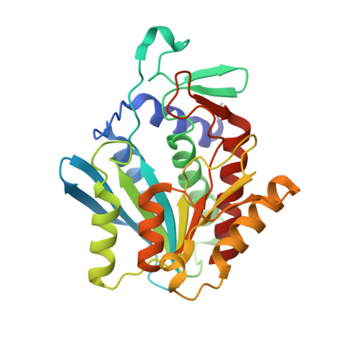
The Dimerization Domain in DapE Enzymes Is required for Catalysis.

Nocek, B.Starus, A.Makowska-Grzyska, M.Gutierrez, B.Sanchez, S.Jedrzejczak, R.Mack, J.C.Olsen, K.W.Joachimiak, A.Holz, R.C.

(2014) PLoS One 9: e93593-e93593

  • DOI: https://doi.org/10.1371/journal.pone.0093593
  • Primary Citation Related Structures: 
    4H2K, 4ONW, 4OP4

  • PubMed Abstract: 

    The emergence of antibiotic-resistant bacterial strains underscores the importance of identifying new drug targets and developing new antimicrobial compounds. Lysine and meso-diaminopimelic acid are essential for protein production and bacterial peptidoglycan cell wall remodeling and are synthesized in bacteria by enzymes encoded within dap operon. Therefore dap enzymes may serve as excellent targets for developing a new class of antimicrobial agents. The dapE-encoded N-succinyl-L,L-diaminopimelic acid desuccinylase (DapE) converts N-succinyl-L,L-diaminopimelic acid to L,L-diaminopimelic acid and succinate. The enzyme is composed of catalytic and dimerization domains, and belongs to the M20 peptidase family. To understand the specific role of each domain of the enzyme we engineered dimerization domain deletion mutants of DapEs from Haemophilus influenzae and Vibrio cholerae, and characterized these proteins structurally and biochemically. No activity was observed for all deletion mutants. Structural comparisons of wild-type, inactive monomeric DapE enzymes with other M20 peptidases suggest that the dimerization domain is essential for DapE enzymatic activity. Structural analysis and molecular dynamics simulations indicate that removal of the dimerization domain increased the flexibility of a conserved active site loop that may provide critical interactions with the substrate.


  • Organizational Affiliation
    • Center for Structural Genomics of Infectious Diseases, Computation Institute, University of Chicago, Chicago, Illinois, United States of America.

Macromolecule Content 

  • Total Structure Weight: 58.39 kDa 
  • Atom Count: 4,697 
  • Modeled Residue Count: 530 
  • Deposited Residue Count: 536 
  • Unique protein chains: 1

Macromolecules

Find similar proteins by:|  3D Structure
Entity ID: 1
MoleculeChains  Sequence LengthOrganismDetailsImage
Succinyl-diaminopimelate desuccinylase
A, B
268Vibrio cholerae O1 biovar El Tor str. N16961Mutation(s): 0 
Gene Names: dapEVC_2152
EC: 3.5.1.18
UniProt
Find proteins for Q9KQ52 (Vibrio cholerae serotype O1 (strain ATCC 39315 / El Tor Inaba N16961))
Explore Q9KQ52 
Go to UniProtKB:  Q9KQ52
Entity Groups
Sequence Clusters30% Identity50% Identity70% Identity90% Identity95% Identity100% Identity
UniProt GroupQ9KQ52
Sequence Annotations
Expand
Reference Sequence

Small Molecules

Ligands 4 Unique
IDChains Name / Formula / InChI Key2D Diagram3D Interactions
GOL

Query on GOL


Download:Ideal Coordinates CCD File
C [auth A],
I [auth B]
GLYCEROL
C3 H8 O3
PEDCQBHIVMGVHV-UHFFFAOYSA-N
BU1

Query on BU1


Download:Ideal Coordinates CCD File
D [auth A]
E [auth A]
H [auth B]
J [auth B]
K [auth B]
D [auth A],
E [auth A],
H [auth B],
J [auth B],
K [auth B],
L [auth B]
1,4-BUTANEDIOL
C4 H10 O2
WERYXYBDKMZEQL-UHFFFAOYSA-N
EDO

Query on EDO


Download:Ideal Coordinates CCD File
F [auth A],
G [auth A],
N [auth B],
O [auth B]
1,2-ETHANEDIOL
C2 H6 O2
LYCAIKOWRPUZTN-UHFFFAOYSA-N
ACT

Query on ACT


Download:Ideal Coordinates CCD File
M [auth B]ACETATE ION
C2 H3 O2
QTBSBXVTEAMEQO-UHFFFAOYSA-M

Experimental Data & Validation

Experimental Data

  • Method: X-RAY DIFFRACTION
  • Resolution: 1.65 Å
  • R-Value Free:  0.171 (Depositor), 0.180 (DCC) 
  • R-Value Work:  0.136 (Depositor), 0.151 (DCC) 
  • R-Value Observed: 0.138 (Depositor) 
Space Group: P 32
Unit Cell:
Length ( Å )Angle ( ˚ )
a = 49.649α = 90
b = 49.649β = 90
c = 232.566γ = 120
Software Package:
Software NamePurpose
SBC-Collectdata collection
CCP4model building
MOLREPphasing
REFMACrefinement
HKL-3000data reduction
HKL-3000data scaling
CCP4phasing

Structure Validation

View Full Validation Report


Entry History 

Deposition Data

Revision History  (Full details and data files)

  • Version 1.0: 2014-04-23
    Type: Initial release
  • Version 1.1: 2014-07-23
    Changes: Database references
  • Version 1.2: 2017-07-26
    Changes: Refinement description, Source and taxonomy
  • Version 1.3: 2023-09-20
    Changes: Data collection, Database references, Derived calculations, Refinement description