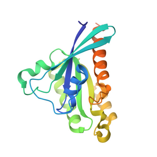
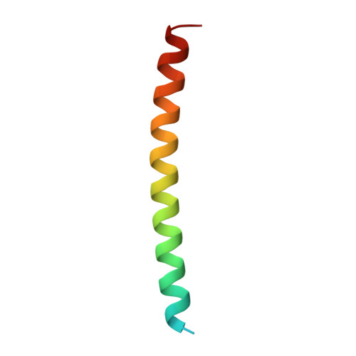
4OJK

Structure of the cGMP Dependent Protein Kinase II and Rab11b Complex


Experimental Data Snapshot

  • Method: X-RAY DIFFRACTION
  • Resolution: 2.66 Å
  • R-Value Free: 0.251 
  • R-Value Work: 0.201 
  • R-Value Observed: 0.204 

Starting Model: experimental
View more details

wwPDB Validation   3D Report Full Report


Ligand Structure Quality Assessment 


This is version 1.4 of the entry. See complete history


Literature

Crystal Structure of the cGMP-dependent Protein Kinase II Leucine Zipper and Rab11b Protein Complex Reveals Molecular Details of G-kinase-specific Interactions.

Reger, A.S.Yang, M.P.Koide-Yoshida, S.Guo, E.Mehta, S.Yuasa, K.Liu, A.Casteel, D.E.Kim, C.

(2014) J Biol Chem 289: 25393-25403

  • DOI: https://doi.org/10.1074/jbc.M114.575894
  • Primary Citation of Related Structures:  
    4OJK

  • PubMed Abstract: 

    cGMP-dependent protein kinase (PKG)-interacting proteins (GKIPs) mediate cellular targeting of PKG isoforms by interacting with their leucine zipper (LZ) domains. These interactions prevent aberrant signaling cross-talk between different PKG isotypes. To gain detailed insight into isotype-specific GKIP recognition by PKG, we analyzed the type II PKG leucine zipper domain and found that residues 40-83 dimerized and specifically interacted with Rab11b. Next, we determined a crystal structure of the PKG II LZ-Rab11b complex. The PKG II LZ domain presents a mostly nonpolar surface onto which Rab11b docks, through van der Waals interactions. Contact surfaces in Rab11b are found in switch I and II, interswitch, and the β1/N-terminal regions. This binding surface dramatically differs from that seen in the Rab11 family of interacting protein complex structures. Structural comparison with PKG Iα and Iβ LZs combined with mutagenic analysis reveals that GKIP recognition is mediated through surface charge interactions.


  • Organizational Affiliation

    From the Department of Pharmacology, Baylor College of Medicine, Houston, Texas 77030.


Macromolecules
Find similar proteins by:  (by identity cutoff)  |  3D Structure
Entity ID: 1
MoleculeChains Sequence LengthOrganismDetailsImage
Ras-related protein Rab-11B
A, B
200Homo sapiensMutation(s): 0 
Gene Names: RAB11BYPT3
EC: 3.6.5.2
UniProt & NIH Common Fund Data Resources
Find proteins for Q15907 (Homo sapiens)
Explore Q15907 
Go to UniProtKB:  Q15907
PHAROS:  Q15907
GTEx:  ENSG00000185236 
Entity Groups  
Sequence Clusters30% Identity50% Identity70% Identity90% Identity95% Identity100% Identity
UniProt GroupQ15907
Sequence Annotations
Expand
  • Reference Sequence
Find similar proteins by:  (by identity cutoff)  |  3D Structure
Entity ID: 2
MoleculeChains Sequence LengthOrganismDetailsImage
cGMP-dependent protein kinase 2
C, D
46Rattus norvegicusMutation(s): 0 
Gene Names: Prkg2
UniProt
Find proteins for Q64595 (Rattus norvegicus)
Explore Q64595 
Go to UniProtKB:  Q64595
Entity Groups  
Sequence Clusters30% Identity50% Identity70% Identity90% Identity95% Identity100% Identity
UniProt GroupQ64595
Sequence Annotations
Expand
  • Reference Sequence
Small Molecules
Experimental Data & Validation

Experimental Data

  • Method: X-RAY DIFFRACTION
  • Resolution: 2.66 Å
  • R-Value Free: 0.251 
  • R-Value Work: 0.201 
  • R-Value Observed: 0.204 
  • Space Group: P 43 21 2
Unit Cell:
Length ( Å )Angle ( ˚ )
a = 136.007α = 90
b = 136.007β = 90
c = 76.997γ = 90
Software Package:
Software NamePurpose
Specdata collection
PHASERphasing
PHENIXrefinement
MOSFLMdata reduction
SCALAdata scaling

Structure Validation

View Full Validation Report


Ligand Structure Quality Assessment 


Entry History 

Deposition Data

Revision History  (Full details and data files)

  • Version 1.0: 2014-08-06
    Type: Initial release
  • Version 1.1: 2014-08-20
    Changes: Derived calculations
  • Version 1.2: 2014-10-01
    Changes: Database references
  • Version 1.3: 2017-11-22
    Changes: Refinement description
  • Version 1.4: 2023-09-20
    Changes: Data collection, Database references, Derived calculations, Refinement description