4OH2 | pdb_00004oh2

Crystal Structure of Cu/Zn Superoxide Dismutase I149T


Experimental Data Snapshot

  • Method: X-RAY DIFFRACTION
  • Resolution: 2.38 Å
  • R-Value Free: 
    0.216 (Depositor), 0.218 (DCC) 
  • R-Value Work: 
    0.163 (Depositor), 0.174 (DCC) 
  • R-Value Observed: 
    0.164 (Depositor) 

wwPDB Validation 3D Report Full Report

Validation slider image for 4OH2

This is version 1.3 of the entry. See complete history

Literature

Copper-Based Pulsed Dipolar ESR Spectroscopy as a Probe of Protein Conformation Linked to Disease States.

Merz, G.E.Borbat, P.P.Pratt, A.J.Getzoff, E.D.Freed, J.H.Crane, B.R.

(2014) Biophys J 107: 1669-1674

  • DOI: https://doi.org/10.1016/j.bpj.2014.07.068
  • Primary Citation Related Structures: 
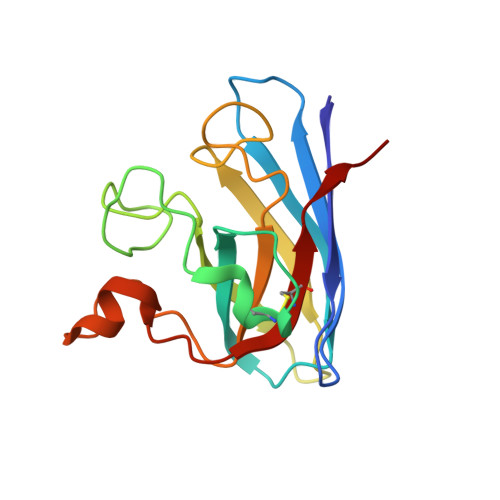
    4OH2

  • PubMed Abstract: 

    We demonstrate the ability of pulsed dipolar electron spin resonance (ESR) spectroscopy (PDS) to report on the conformation of Cu-Zn superoxide dismutase (SOD1) through the sensitive measurement of dipolar interactions between inherent Cu(2+) ions. Although the extent and the anisotropy of the Cu ESR spectrum provides challenges for PDS, Ku-band (17.3 GHz) double electron-electron resonance and double-quantum coherence variants of PDS coupled with distance reconstruction methods recover Cu-Cu distances in good agreement with crystal structures. Moreover, Cu-PDS measurements expose distinct differences between the conformational properties of wild-type SOD1 and a single-residue variant (I149T) that leads to the disease amyotrophic lateral sclerosis (ALS). The I149T protein displays a broader Cu-Cu distance distribution within the SOD1 dimer compared to wild-type. In a nitroxide (NO)-labeled sample, distance distributions obtained from Cu-Cu, Cu-NO, and NO-NO separations reveal increased structural heterogeneity within the protein and a tendency for mutant dimers to associate. In contrast, perturbations caused by the ALS mutation are completely masked in the crystal structure of I149T. Thus, PDS readily detects alterations in metalloenzyme solution properties not easily deciphered by other methods and in doing so supports the notion that increased range of motion and associations of SOD1 ALS variants contribute to disease progression.


  • Organizational Affiliation
    • Department of Chemistry and Chemical Biology, Cornell University, Ithaca, New York.

Macromolecule Content 

  • Total Structure Weight: 158.96 kDa 
  • Atom Count: 11,814 
  • Modeled Residue Count: 1,530 
  • Deposited Residue Count: 1,530 
  • Unique protein chains: 1

Macromolecules

Find similar proteins by:|  3D Structure
Entity ID: 1
MoleculeChains  Sequence LengthOrganismDetailsImage
Superoxide dismutase [Cu-Zn]
A, B, C, D, E
A, B, C, D, E, F, G, H, I, J
153Homo sapiensMutation(s): 3 
Gene Names: Homo sapiens SOD1SOD1
EC: 1.15.1.1 (PDB Primary Data), 1.8 (UniProt)
UniProt & NIH Common Fund Data Resources
Find proteins for P00441 (Homo sapiens)
Explore P00441 
Go to UniProtKB:  P00441
PHAROS:  P00441
GTEx:  ENSG00000142168 
Entity Groups
Sequence Clusters30% Identity50% Identity70% Identity90% Identity95% Identity100% Identity
UniProt GroupP00441
Sequence Annotations
Expand
Reference Sequence

Small Molecules

Ligands 2 Unique
IDChains Name / Formula / InChI Key2D Diagram3D Interactions
ZN

Query on ZN


Download:Ideal Coordinates CCD File
BA [auth I]
DA [auth J]
L [auth A]
N [auth B]
P [auth C]
BA [auth I],
DA [auth J],
L [auth A],
N [auth B],
P [auth C],
R [auth D],
T [auth E],
V [auth F],
X [auth G],
Z [auth H]
ZINC ION
Zn
PTFCDOFLOPIGGS-UHFFFAOYSA-N
CU

Query on CU


Download:Ideal Coordinates CCD File
AA [auth I]
CA [auth J]
K [auth A]
M [auth B]
O [auth C]
AA [auth I],
CA [auth J],
K [auth A],
M [auth B],
O [auth C],
Q [auth D],
S [auth E],
U [auth F],
W [auth G],
Y [auth H]
COPPER (II) ION
Cu
JPVYNHNXODAKFH-UHFFFAOYSA-N

Experimental Data & Validation

Experimental Data

  • Method: X-RAY DIFFRACTION
  • Resolution: 2.38 Å
  • R-Value Free:  0.216 (Depositor), 0.218 (DCC) 
  • R-Value Work:  0.163 (Depositor), 0.174 (DCC) 
  • R-Value Observed: 0.164 (Depositor) 
Space Group: C 2 2 21
Unit Cell:
Length ( Å )Angle ( ˚ )
a = 165.099α = 90
b = 203.811β = 90
c = 144.407γ = 90
Software Package:
Software NamePurpose
DENZOdata reduction
SCALEPACKdata scaling
PHASERphasing
PHENIXrefinement
PDB_EXTRACTdata extraction

Structure Validation

View Full Validation Report


Entry History 

Deposition Data

Revision History  (Full details and data files)

  • Version 1.0: 2014-10-15
    Type: Initial release
  • Version 1.1: 2014-10-22
    Changes: Database references
  • Version 1.2: 2017-11-22
    Changes: Advisory, Refinement description
  • Version 1.3: 2024-11-27
    Changes: Advisory, Data collection, Database references, Derived calculations, Structure summary