Structural and Evolutionary Analyses Show Unique Stabilization Strategies in the Type IV Pili of Clostridium difficile.
Piepenbrink, K.H., Maldarelli, G.A., Martinez de la Pena, C.F., Dingle, T.C., Mulvey, G.L., Lee, A., von Rosenvinge, E., Armstrong, G.D., Donnenberg, M.S., Sundberg, E.J.(2015) Structure 23: 385-396
- PubMed: 25599642 Search on PubMedSearch on PubMed Central
- DOI: https://doi.org/10.1016/j.str.2014.11.018
- Primary Citation Related Structures:
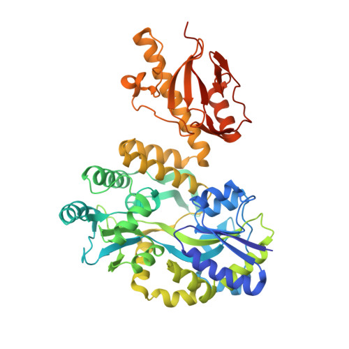
4OGM, 4PE2, 4TSM - PubMed Abstract:
Type IV pili are produced by many pathogenic Gram-negative bacteria and are important for processes as diverse as twitching motility, biofilm formation, cellular adhesion, and horizontal gene transfer. However, many Gram-positive species, including Clostridium difficile, also produce type IV pili. Here, we identify the major subunit of the type IV pili of C. difficile, PilA1, and describe multiple 3D structures of PilA1, demonstrating the diversity found in three strains of C. difficile. We also model the incorporation of both PilA1 and a minor pilin, PilJ, into the pilus fiber. Although PilA1 contains no cysteine residues, and therefore cannot form the disulfide bonds found in all Gram-negative type IV pilins, it adopts unique strategies to achieve a typical pilin fold. The structures of PilA1 and PilJ exhibit similarities with the type IVb pilins from Gram-negative bacteria that suggest that the type IV pili of C. difficile are involved in microcolony formation.
- Institute of Human Virology, University of Maryland School of Medicine, Baltimore, MD 21201, USA.
Organizational Affiliation: