4O96 | pdb_00004o96

2.60 Angstrom resolution crystal structure of a protein kinase domain of type III effector NleH2 (ECs1814) from Escherichia coli O157:H7 str. Sakai


Experimental Data Snapshot

  • Method: X-RAY DIFFRACTION
  • Resolution: 2.60 Å
  • R-Value Free: 
    0.253 (Depositor), 0.247 (DCC) 
  • R-Value Work: 
    0.208 (Depositor) 
  • R-Value Observed: 
    0.210 (Depositor) 

wwPDB Validation 3D Report Full Report

Validation slider image for 4O96

This is version 1.4 of the entry. See complete history

Literature

Type III Effector NleH2 from Escherichia coli O157:H7 str. Sakai Features an Atypical Protein Kinase Domain.

Halavaty, A.S.Anderson, S.M.Wawrzak, Z.Kudritska, M.Skarina, T.Anderson, W.F.Savchenko, A.

(2014) Biochemistry 53: 2433-2435

  • DOI: https://doi.org/10.1021/bi500016j
  • Primary Citation Related Structures: 
    4O96

  • PubMed Abstract: 

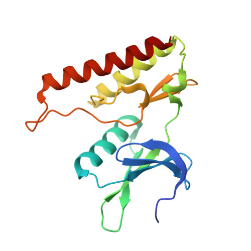
    The crystal structure of a C-terminal domain of enterohemorrhagic Escherichia coli type III effector NleH2 has been determined to 2.6 Å resolution. The structure resembles those of protein kinases featuring the catalytic, activation, and glycine-rich loop motifs and ATP-binding site. The position of helix αC and the lack of a conserved arginine within an equivalent HRD motif suggested that the NleH2 kinase domain's active conformation might not require phosphorylation. The activation segment markedly contributed to the dimerization interface of NleH2, which can also accommodate the NleH1-NleH2 heterodimer. The C-terminal PDZ-binding motif of NleH2 provided bases for interaction with host proteins.


  • Organizational Affiliation
    • Center for Structural Genomics of Infectious Diseases (CSGID), Molecular Pharmacology and Biological Chemistry, Northwestern University , Chicago, Illinois 60611, United States.

Macromolecule Content 

  • Total Structure Weight: 74.86 kDa 
  • Atom Count: 5,400 
  • Modeled Residue Count: 652 
  • Deposited Residue Count: 664 
  • Unique protein chains: 1

Macromolecules

Find similar proteins by:|  3D Structure
Entity ID: 1
MoleculeChains  Sequence LengthOrganismDetailsImage
type III effector protein kinase
A, B, C, D
166Escherichia coli O157:H7 str. SakaiMutation(s): 0 
Gene Names: ECs1814Z6021
UniProt
Find proteins for Q8XAL6 (Escherichia coli O157:H7)
Explore Q8XAL6 
Go to UniProtKB:  Q8XAL6
Entity Groups
Sequence Clusters30% Identity50% Identity70% Identity90% Identity95% Identity100% Identity
UniProt GroupQ8XAL6
Sequence Annotations
Expand
Reference Sequence

Experimental Data & Validation

Experimental Data

  • Method: X-RAY DIFFRACTION
  • Resolution: 2.60 Å
  • R-Value Free:  0.253 (Depositor), 0.247 (DCC) 
  • R-Value Work:  0.208 (Depositor) 
  • R-Value Observed: 0.210 (Depositor) 
Space Group: P 4 21 2
Unit Cell:
Length ( Å )Angle ( ˚ )
a = 147.252α = 90
b = 147.252β = 90
c = 83.05γ = 90
Software Package:
Software NamePurpose
Blu-Icedata collection
PHENIXmodel building
REFMACrefinement
HKL-2000data reduction
HKL-2000data scaling
PHENIXphasing

Structure Validation

View Full Validation Report


Entry History 

Revision History  (Full details and data files)

  • Version 1.0: 2014-01-15
    Type: Initial release
  • Version 1.1: 2014-04-16
    Changes: Database references, Structure summary
  • Version 1.2: 2014-05-21
    Changes: Database references
  • Version 1.3: 2017-11-22
    Changes: Refinement description
  • Version 1.4: 2024-02-28
    Changes: Data collection, Database references, Derived calculations, Refinement description