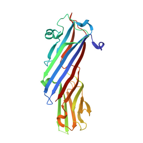
4O8Q

Crystal structure of bovine MHD domain of the COPI delta subunit at 2.15 A resolution


Experimental Data Snapshot

  • Method: X-RAY DIFFRACTION
  • Resolution: 2.15 Å
  • R-Value Free: 0.229 
  • R-Value Work: 0.182 
  • R-Value Observed: 0.184 

Starting Model: experimental
View more details

wwPDB Validation   3D Report Full Report


This is version 1.4 of the entry. See complete history


Literature

Structure of the bovine COPI delta subunit mu homology domain at 2.15 angstrom resolution.

Lahav, A.Rozenberg, H.Parnis, A.Cassel, D.Adir, N.

(2015) Acta Crystallogr D Biol Crystallogr 71: 1328-1334

  • DOI: https://doi.org/10.1107/S1399004715006203
  • Primary Citation of Related Structures:  
    4O8Q

  • PubMed Abstract: 

    The heptameric COPI coat (coatomer) plays an essential role in vesicular transport in the early secretory system of eukaryotic cells. While the structures of some of the subunits have been determined, that of the δ-COP subunit has not been reported to date. The δ-COP subunit is part of a subcomplex with structural similarity to tetrameric clathrin adaptors (APs), where δ-COP is the structural homologue of the AP μ subunit. Here, the crystal structure of the μ homology domain (MHD) of δ-COP (δ-MHD) obtained by phasing using a combined SAD-MR method is presented at 2.15 Å resolution. The crystallographic asymmetric unit contains two monomers that exhibit short sections of disorder, which may allude to flexible regions of the protein. The δ-MHD is composed of two subdomains connected by unstructured linkers. Comparison between this structure and those of known MHD domains from the APs shows significant differences in the positions of specific loops and β-sheets, as well as a more general change in the relative positions of the protein subdomains. The identified difference may be the major source of cargo-binding specificity. Finally, the crystal structure is used to analyze the potential effect of the I422T mutation in δ-COP previously reported to cause a neurodegenerative phenotype in mice.


  • Organizational Affiliation

    Schulich Faculty of Chemistry, Technion, Haifa 32000, Israel.


Macromolecules
Find similar proteins by:  (by identity cutoff)  |  3D Structure
Entity ID: 1
MoleculeChains Sequence LengthOrganismDetailsImage
Coatomer subunit delta
A, B
245Bos taurusMutation(s): 0 
Gene Names: ARCN1COPD
UniProt
Find proteins for P53619 (Bos taurus)
Explore P53619 
Go to UniProtKB:  P53619
Entity Groups  
Sequence Clusters30% Identity50% Identity70% Identity90% Identity95% Identity100% Identity
UniProt GroupP53619
Sequence Annotations
Expand
  • Reference Sequence
Small Molecules
Ligands 1 Unique
IDChains Name / Formula / InChI Key2D Diagram3D Interactions
FMT
Query on FMT

Download Ideal Coordinates CCD File 
C [auth A]FORMIC ACID
C H2 O2
BDAGIHXWWSANSR-UHFFFAOYSA-N
Experimental Data & Validation

Experimental Data

  • Method: X-RAY DIFFRACTION
  • Resolution: 2.15 Å
  • R-Value Free: 0.229 
  • R-Value Work: 0.182 
  • R-Value Observed: 0.184 
  • Space Group: P 21 21 21
Unit Cell:
Length ( Å )Angle ( ˚ )
a = 42.092α = 90
b = 110.333β = 90
c = 145.794γ = 90
Software Package:
Software NamePurpose
DNAdata collection
PHASERphasing
PHENIXrefinement
HKL-2000data reduction
HKL-2000data scaling

Structure Validation

View Full Validation Report


Entry History 

Deposition Data

Revision History  (Full details and data files)

  • Version 1.0: 2015-01-07
    Type: Initial release
  • Version 1.1: 2015-06-10
    Changes: Database references
  • Version 1.2: 2015-06-24
    Changes: Database references
  • Version 1.3: 2024-02-28
    Changes: Data collection, Database references, Derived calculations
  • Version 1.4: 2024-04-03
    Changes: Refinement description