Elucidation of the molecular basis for arabinoxylan-debranching activity of a thermostable family GH62 alpha-l-arabinofuranosidase from Streptomyces thermoviolaceus.
Wang, W., Mai-Gisondi, G., Stogios, P.J., Kaur, A., Xu, X., Cui, H., Turunen, O., Savchenko, A., Master, E.R.(2014) Appl Environ Microbiol 80: 5317-5329
- PubMed: 24951792 Search on PubMedSearch on PubMed Central
- DOI: https://doi.org/10.1128/AEM.00685-14
- Primary Citation Related Structures:
4O8N, 4O8O, 4O8P - PubMed Abstract:
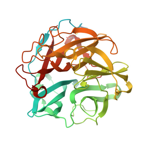
Xylan-debranching enzymes facilitate the complete hydrolysis of xylan and can be used to alter xylan chemistry. Here, the family GH62 α-l-arabinofuranosidase from Streptomyces thermoviolaceus (SthAbf62A) was shown to have a half-life of 60 min at 60°C and the ability to cleave α-1,3 l-arabinofuranose (l-Araf) from singly substituted xylopyranosyl (Xylp) backbone residues in wheat arabinoxylan; low levels of activity on arabinan as well as 4-nitrophenyl α-l-arabinofuranoside were also detected. After selective removal of α-1,3 l-Araf substituents from disubstituted Xylp residues present in wheat arabinoxylan, SthAbf62A could also cleave the remaining α-1,2 l-Araf substituents, confirming the ability of SthAbf62A to remove α-l-Araf residues that are (1→2) and (1→3) linked to monosubstituted β-d-Xylp sugars. Three-dimensional structures of SthAbf62A and its complex with xylotetraose and l-arabinose confirmed a five-bladed β-propeller fold and revealed a molecular Velcro in blade V between the β1 and β21 strands, a disulfide bond between Cys27 and Cys297, and a calcium ion coordinated in the central channel of the fold. The enzyme-arabinose complex structure further revealed a narrow and seemingly rigid l-arabinose binding pocket situated at the center of one side of the β propeller, which stabilized the arabinofuranosyl substituent through several hydrogen-bonding and hydrophobic interactions. The predicted catalytic amino acids were oriented toward this binding pocket, and the catalytic essentiality of Asp53 and Glu213 was confirmed by site-specific mutagenesis. Complex structures with xylotetraose revealed a shallow cleft for xylan backbone binding that is open at both ends and comprises multiple binding subsites above and flanking the l-arabinose binding pocket.
- Department of Chemical Engineering and Applied Chemistry, University of Toronto, Toronto, Ontario, Canada.
Organizational Affiliation: