4O8A

First structure of a proline utilization A proline dehydrogenase domain


Experimental Data Snapshot

  • Method: X-RAY DIFFRACTION
  • Resolution: 2.00 Å
  • R-Value Free: 0.219 
  • R-Value Work: 0.186 
  • R-Value Observed: 0.189 

wwPDB Validation   3D Report Full Report


Ligand Structure Quality Assessment 


This is version 1.1 of the entry. See complete history


Literature

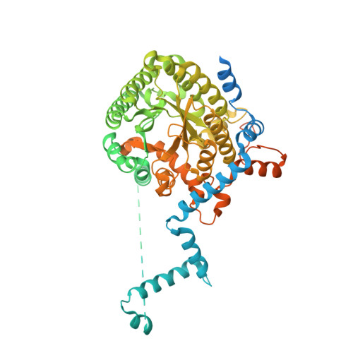
Structure of the proline dehydrogenase domain of the multifunctional PutA flavoprotein.

Lee, Y.H.Nadaraia, S.Gu, D.Becker, D.F.Tanner, J.J.

(2003) Nat Struct Biol 10: 109-114

  • DOI: https://doi.org/10.1038/nsb885
  • Primary Citation of Related Structures:  
    4O8A

  • PubMed Abstract: 

    The PutA flavoprotein from Escherichia coli plays multiple roles in proline catabolism by functioning as a membrane-associated bi-functional enzyme and a transcriptional repressor of proline utilization genes. The human homolog of the PutA proline dehydrogenase (PRODH) domain is critical in p53-mediated apoptosis and schizophrenia. Here we report the crystal structure of a 669-residue truncated form of PutA that shows both PRODH and DNA-binding activities, representing the first structure of a PutA protein and a PRODH enzyme from any organism. The structure is a domain-swapped dimer with each subunit comprising three domains: a helical dimerization arm, a 120-residue domain containing a three-helix bundle similar to that in the helix-turn-helix superfamily of DNA-binding proteins and a beta/alpha-barrel PRODH domain with a bound lactate inhibitor. Analysis of the structure provides insight into the mechanism of proline oxidation to pyrroline-5-carboxylate, and functional studies of a mutant protein suggest that the DNA-binding domain is located within the N-terminal 261 residues of E. coli PutA.


  • Organizational Affiliation

    Department of Chemistry and Biochemistry, University of Missouri-Columbia, 65211, USA.


Macromolecules
Find similar proteins by:  (by identity cutoff)  |  3D Structure
Entity ID: 1
MoleculeChains Sequence LengthOrganismDetailsImage
Bifunctional protein PutA687Escherichia coli K-12Mutation(s): 0 
Gene Names: b1014JW0999poaAputA
EC: 1.5.99.8 (PDB Primary Data), 1.2.1.88 (PDB Primary Data)
UniProt
Find proteins for P09546 (Escherichia coli (strain K12))
Explore P09546 
Go to UniProtKB:  P09546
Entity Groups  
Sequence Clusters30% Identity50% Identity70% Identity90% Identity95% Identity100% Identity
UniProt GroupP09546
Sequence Annotations
Expand
  • Reference Sequence
Experimental Data & Validation

Experimental Data

  • Method: X-RAY DIFFRACTION
  • Resolution: 2.00 Å
  • R-Value Free: 0.219 
  • R-Value Work: 0.186 
  • R-Value Observed: 0.189 
  • Space Group: I 2 2 2
Unit Cell:
Length ( Å )Angle ( ˚ )
a = 72.62α = 90
b = 140.43β = 90
c = 145.85γ = 90
Software Package:
Software NamePurpose
PHENIXrefinement
PDB_EXTRACTdata extraction
DENZOdata reduction
SCALEPACKdata scaling
MLPHAREphasing

Structure Validation

View Full Validation Report


Ligand Structure Quality Assessment 


Entry History 

Deposition Data

  • Released Date: 2014-01-15 
  • Deposition Author(s): Tanner, J.J.
  • This entry supersedes: 1K87

Revision History  (Full details and data files)

  • Version 1.0: 2014-01-15
    Type: Initial release
  • Version 1.1: 2024-02-28
    Changes: Data collection, Database references, Derived calculations