A transcription blocker isolated from a designed repeat protein combinatorial library by in vivo functional screen.
Tikhonova, E.B., Ethayathulla, A.S., Su, Y., Hariharan, P., Xie, S., Guan, L.(2015) Sci Rep 5: 8070-8070
- PubMed: 25627011 Search on PubMedSearch on PubMed Central
- DOI: https://doi.org/10.1038/srep08070
- Primary Citation Related Structures:
4O60, 4QFV - PubMed Abstract:
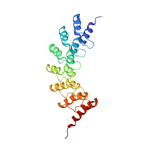
A highly diverse DNA library coding for ankyrin seven-repeat proteins (ANK-N5C) was designed and constructed by a PCR-based combinatorial assembly strategy. A bacterial melibiose fermentation assay was adapted for in vivo functional screen. We isolated a transcription blocker that completely inhibits the melibiose-dependent expression of α-galactosidase (MelA) and melibiose permease (MelB) of Escherichia coli by specifically preventing activation of the melAB operon. High-resolution crystal structural determination reveals that the designed ANK-N5C protein has a typical ankyrin fold, and the specific transcription blocker, ANK-N5C-281, forms a domain-swapped dimer. Functional tests suggest that the activity of MelR, a DNA-binding transcription activator and a member of AraC family of transcription factors, is inhibited by ANK-N5C-281 protein. All ANK-N5C proteins are expected to have a concave binding area with negative surface potential, suggesting that the designed ANK-N5C library proteins may facilitate the discovery of binders recognizing structural motifs with positive surface potential, like in DNA-binding proteins. Overall, our results show that the established library is a useful tool for the discovery of novel bioactive reagents.
- Department of Cell Physiology &Molecular Biophysics, Center for Membrane Protein Research, Texas Tech University Health Sciences Center, Lubbock, Texas 79430.
Organizational Affiliation: