4NZU | pdb_00004nzu

Crystal structure of the primary monoclonal antibody 13PL Fab' from a multiple myeloma patient


Experimental Data Snapshot

  • Method: X-RAY DIFFRACTION
  • Resolution: 1.20 Å
  • R-Value Free: 
    0.163 (Depositor), 0.160 (DCC) 
  • R-Value Work: 
    0.136 (Depositor) 
  • R-Value Observed: 
    0.137 (Depositor) 

Starting Model: experimental
View more details

wwPDB Validation 3D Report Full Report

Validation slider image for 4NZU

This is version 1.2 of the entry. See complete history

Literature

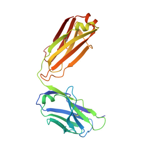
A structurally distinct human mycoplasma protein that generically blocks antigen-antibody union.

Grover, R.K.Zhu, X.Nieusma, T.Jones, T.Boero, I.MacLeod, A.S.Mark, A.Niessen, S.Kim, H.J.Kong, L.Assad-Garcia, N.Kwon, K.Chesi, M.Smider, V.V.Salomon, D.R.Jelinek, D.F.Kyle, R.A.Pyles, R.B.Glass, J.I.Ward, A.B.Wilson, I.A.Lerner, R.A.

(2014) Science 343: 656-661

  • DOI: https://doi.org/10.1126/science.1246135
  • Primary Citation Related Structures: 
    4NZR, 4NZT, 4NZU

  • PubMed Abstract: 

    We report the discovery of a broadly reactive antibody-binding protein (Protein M) from human mycoplasma. The crystal structure of the ectodomain of transmembrane Protein M differs from other known protein structures, as does its mechanism of antibody binding. Protein M binds with high affinity to all types of human and nonhuman immunoglobulin G, predominantly through attachment to the conserved portions of the variable region of the κ and λ light chains. Protein M blocks antibody-antigen union, likely because of its large C-terminal domain extending over the antibody-combining site, blocking entry to large antigens. Similar to the other immunoglobulin-binding proteins such as Protein A, Protein M as well as its orthologs in other Mycoplasma species could become invaluable reagents in the antibody field.


  • Organizational Affiliation
    • Department of Cell and Molecular Biology, The Scripps Research Institute, La Jolla, CA 92037, USA.

Macromolecule Content 

  • Total Structure Weight: 47.08 kDa 
  • Atom Count: 3,918 
  • Modeled Residue Count: 434 
  • Deposited Residue Count: 436 
  • Unique protein chains: 2

Macromolecules

Find similar proteins by:|  3D Structure
Entity ID: 1
MoleculeChains  Sequence LengthOrganismDetailsImage
13PL heavy chainA [auth L]211Homo sapiensMutation(s): 0 
Find similar proteins by:|  3D Structure
Entity ID: 2
MoleculeChains  Sequence LengthOrganismDetailsImage
13PL light chainB [auth H]225Homo sapiensMutation(s): 0 

Experimental Data & Validation

Experimental Data

  • Method: X-RAY DIFFRACTION
  • Resolution: 1.20 Å
  • R-Value Free:  0.163 (Depositor), 0.160 (DCC) 
  • R-Value Work:  0.136 (Depositor) 
  • R-Value Observed: 0.137 (Depositor) 
Space Group: P 21 21 21
Unit Cell:
Length ( Å )Angle ( ˚ )
a = 51.179α = 90
b = 80.403β = 90
c = 102.607γ = 90
Software Package:
Software NamePurpose
HKL-2000data collection
PHASERphasing
REFMACrefinement
HKL-2000data reduction
HKL-2000data scaling

Structure Validation

View Full Validation Report


Entry History 

Deposition Data

Revision History  (Full details and data files)

  • Version 1.0: 2014-02-19
    Type: Initial release
  • Version 1.1: 2023-09-20
    Changes: Data collection, Database references, Derived calculations, Refinement description
  • Version 1.2: 2024-10-30
    Changes: Structure summary