4NZM | pdb_00004nzm

Crystal structure of the catalytic domain of PPIP5K2 in complex with AMPPNP and 5-PA-InsP5


Experimental Data Snapshot

  • Method: X-RAY DIFFRACTION
  • Resolution: 2.00 Å
  • R-Value Free: 
    0.231 (Depositor), 0.233 (DCC) 
  • R-Value Work: 
    0.198 (Depositor), 0.199 (DCC) 
  • R-Value Observed: 
    0.200 (Depositor) 

Starting Model: experimental
View more details

wwPDB Validation 3D Report Full Report

Validation slider image for 4NZM

Ligand Structure Quality Assessment 


This is version 1.2 of the entry. See complete history

Literature

Synthetic Inositol Phosphate Analogs Reveal that PPIP5K2 Has a Surface-Mounted Substrate Capture Site that Is a Target for Drug Discovery.

Wang, H.Godage, H.Y.Riley, A.M.Weaver, J.D.Shears, S.B.Potter, B.V.

(2014) Chem Biol 21: 689-699

  • DOI: https://doi.org/10.1016/j.chembiol.2014.03.009
  • Primary Citation Related Structures: 
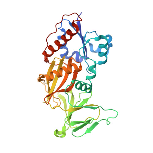
    4NZM, 4NZN, 4NZO

  • PubMed Abstract: 

    Diphosphoinositol pentakisphosphate kinase 2 (PPIP5K2) is one of the mammalian PPIP5K isoforms responsible for synthesis of diphosphoinositol polyphosphates (inositol pyrophosphates; PP-InsPs), regulatory molecules that function at the interface of cell signaling and organismic homeostasis. The development of drugs that inhibit PPIP5K2 could have both experimental and therapeutic applications. Here, we describe a synthetic strategy for producing naturally occurring 5-PP-InsP4, as well as several inositol polyphosphate analogs, and we study their interactions with PPIP5K2 using biochemical and structural approaches. These experiments uncover an additional ligand-binding site on the surface of PPIP5K2, adjacent to the catalytic pocket. This site facilitates substrate capture from the bulk phase, prior to transfer into the catalytic pocket. In addition to demonstrating a "catch-and-pass" reaction mechanism in a small molecule kinase, we demonstrate that binding of our analogs to the substrate capture site inhibits PPIP5K2. This work suggests that the substrate-binding site offers new opportunities for targeted drug design.


  • Organizational Affiliation
    • Inositol Signaling Group, Laboratory of Signal Transduction, National Institute of Environmental Health Sciences, National Institutes of Health, Research Triangle Park, NC 27709, USA.

Macromolecule Content 

  • Total Structure Weight: 38.87 kDa 
  • Atom Count: 3,161 
  • Modeled Residue Count: 325 
  • Deposited Residue Count: 330 
  • Unique protein chains: 1

Macromolecules

Find similar proteins by:|  3D Structure
Entity ID: 1
MoleculeChains  Sequence LengthOrganismDetailsImage
Inositol hexakisphosphate and diphosphoinositol-pentakisphosphate kinase 2330Homo sapiensMutation(s): 0 
Gene Names: PPIP5K2HISPPD1KIAA0433VIP2
EC: 2.7.4.21 (PDB Primary Data), 2.7.4.24 (PDB Primary Data)
UniProt & NIH Common Fund Data Resources
Find proteins for O43314 (Homo sapiens)
Explore O43314 
Go to UniProtKB:  O43314
PHAROS:  O43314
GTEx:  ENSG00000145725 
Entity Groups
Sequence Clusters30% Identity50% Identity70% Identity90% Identity95% Identity100% Identity
UniProt GroupO43314
Sequence Annotations
Expand
Reference Sequence

Small Molecules

Ligands 3 Unique
IDChains Name / Formula / InChI Key2D Diagram3D Interactions
0EJ

Query on 0EJ


Download:Ideal Coordinates CCD File
C [auth A](2-oxo-2-{[(1s,2R,3S,4s,5R,6S)-2,3,4,5,6-pentakis(phosphonooxy)cyclohexyl]oxy}ethyl)phosphonic acid
C8 H20 O25 P6
JGWVNODNEALCIO-QEEWUKHFSA-N
ANP

Query on ANP


Download:Ideal Coordinates CCD File
B [auth A]PHOSPHOAMINOPHOSPHONIC ACID-ADENYLATE ESTER
C10 H17 N6 O12 P3
PVKSNHVPLWYQGJ-KQYNXXCUSA-N
MG

Query on MG


Download:Ideal Coordinates CCD File
D [auth A],
E [auth A],
F [auth A],
G [auth A]
MAGNESIUM ION
Mg
JLVVSXFLKOJNIY-UHFFFAOYSA-N

Experimental Data & Validation

Experimental Data

  • Method: X-RAY DIFFRACTION
  • Resolution: 2.00 Å
  • R-Value Free:  0.231 (Depositor), 0.233 (DCC) 
  • R-Value Work:  0.198 (Depositor), 0.199 (DCC) 
  • R-Value Observed: 0.200 (Depositor) 
Space Group: P 21 21 21
Unit Cell:
Length ( Å )Angle ( ˚ )
a = 89.623α = 90
b = 111.226β = 90
c = 41.443γ = 90
Software Package:
Software NamePurpose
HKL-2000data collection
CCP4model building
REFMACrefinement
HKL-2000data reduction
HKL-2000data scaling
CCP4phasing

Structure Validation

View Full Validation Report


Ligand Structure Quality Assessment 


Entry History 

Deposition Data

Revision History  (Full details and data files)

  • Version 1.0: 2014-04-02
    Type: Initial release
  • Version 1.1: 2014-06-11
    Changes: Database references
  • Version 1.2: 2023-09-20
    Changes: Data collection, Database references, Derived calculations, Refinement description