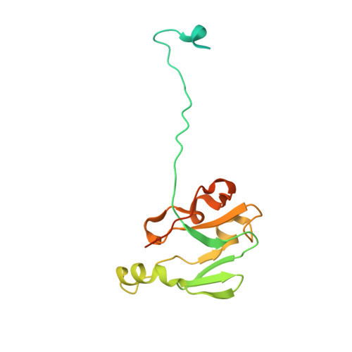
1.8 Angstrom Crystal Structure of Signal Peptidase I from Bacillus anthracis.
Minasov, G., Shuvalova, L., Dubrovska, I., Winsor, J., Shatsman, S., Kwon, K., Anderson, W.F., Center for Structural Genomics of Infectious Diseases (CSGID)To be published.