4NTW

Structure of acid-sensing ion channel in complex with snake toxin


Experimental Data Snapshot

  • Method: X-RAY DIFFRACTION
  • Resolution: 2.07 Å
  • R-Value Free: 0.246 
  • R-Value Work: 0.206 
  • R-Value Observed: 0.208 

wwPDB Validation   3D Report Full Report


This is version 2.1 of the entry. See complete history


Literature

X-ray structure of Acid-sensing ion channel 1-snake toxin complex reveals open state of a na(+)-selective channel.

Baconguis, I.Bohlen, C.J.Goehring, A.Julius, D.Gouaux, E.

(2014) Cell 156: 717-729

  • DOI: https://doi.org/10.1016/j.cell.2014.01.011
  • Primary Citation of Related Structures:  
    4NTW, 4NTX, 4NTY

  • PubMed Abstract: 

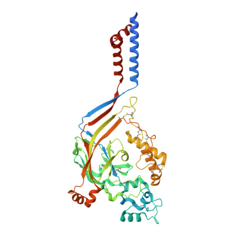
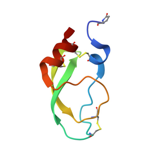
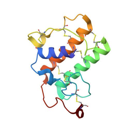
    Acid-sensing ion channels (ASICs) detect extracellular protons produced during inflammation or ischemic injury and belong to the superfamily of degenerin/epithelial sodium channels. Here, we determine the cocrystal structure of chicken ASIC1a with MitTx, a pain-inducing toxin from the Texas coral snake, to define the structure of the open state of ASIC1a. In the MitTx-bound open state and in the previously determined low-pH desensitized state, TM2 is a discontinuous α helix in which the Gly-Ala-Ser selectivity filter adopts an extended, belt-like conformation, swapping the cytoplasmic one-third of TM2 with an adjacent subunit. Gly 443 residues of the selectivity filter provide a ring of three carbonyl oxygen atoms with a radius of ∼3.6 Å, presenting an energetic barrier for hydrated ions. The ASIC1a-MitTx complex illuminates the mechanism of MitTx action, defines the structure of the selectivity filter of voltage-independent, sodium-selective ion channels, and captures the open state of an ASIC.


  • Organizational Affiliation

    Department of Physiology, University of California, San Francisco, San Francisco, CA 94158, USA.


Macromolecules
Find similar proteins by:  (by identity cutoff)  |  3D Structure
Entity ID: 1
MoleculeChains Sequence LengthOrganismDetailsImage
Acid-sensing ion channel 1450Gallus gallusMutation(s): 0 
Gene Names: ASIC1ACCN2
Membrane Entity: Yes 
UniProt
Find proteins for Q1XA76 (Gallus gallus)
Explore Q1XA76 
Go to UniProtKB:  Q1XA76
Entity Groups  
Sequence Clusters30% Identity50% Identity70% Identity90% Identity95% Identity100% Identity
UniProt GroupQ1XA76
Sequence Annotations
Expand
  • Reference Sequence
Find similar proteins by:  (by identity cutoff)  |  3D Structure
Entity ID: 2
MoleculeChains Sequence LengthOrganismDetailsImage
Neurotoxin MitTx-alpha60Micrurus tener tenerMutation(s): 0 
Membrane Entity: Yes 
UniProt
Find proteins for G9I929 (Micrurus tener tener)
Explore G9I929 
Go to UniProtKB:  G9I929
Entity Groups  
Sequence Clusters30% Identity50% Identity70% Identity90% Identity95% Identity100% Identity
UniProt GroupG9I929
Sequence Annotations
Expand
  • Reference Sequence
Find similar proteins by:  (by identity cutoff)  |  3D Structure
Entity ID: 3
MoleculeChains Sequence LengthOrganismDetailsImage
Basic phospholipase A2 homolog Tx-beta119Micrurus tener tenerMutation(s): 0 
Membrane Entity: Yes 
UniProt
Find proteins for G9I930 (Micrurus tener tener)
Explore G9I930 
Go to UniProtKB:  G9I930
Entity Groups  
Sequence Clusters30% Identity50% Identity70% Identity90% Identity95% Identity100% Identity
UniProt GroupG9I930
Sequence Annotations
Expand
  • Reference Sequence
Small Molecules
Ligands 4 Unique
IDChains Name / Formula / InChI Key2D Diagram3D Interactions
P6G
Query on P6G

Download Ideal Coordinates CCD File 
I [auth A]HEXAETHYLENE GLYCOL
C12 H26 O7
IIRDTKBZINWQAW-UHFFFAOYSA-N
NAG
Query on NAG

Download Ideal Coordinates CCD File 
D [auth A],
E [auth A]
2-acetamido-2-deoxy-beta-D-glucopyranose
C8 H15 N O6
OVRNDRQMDRJTHS-FMDGEEDCSA-N
CL
Query on CL

Download Ideal Coordinates CCD File 
F [auth A]CHLORIDE ION
Cl
VEXZGXHMUGYJMC-UHFFFAOYSA-M
NA
Query on NA

Download Ideal Coordinates CCD File 
G [auth A],
H [auth A],
J [auth C]
SODIUM ION
Na
FKNQFGJONOIPTF-UHFFFAOYSA-N
Modified Residues  1 Unique
IDChains TypeFormula2D DiagramParent
PCA
Query on PCA
B
L-PEPTIDE LINKINGC5 H7 N O3GLN
Experimental Data & Validation

Experimental Data

  • Method: X-RAY DIFFRACTION
  • Resolution: 2.07 Å
  • R-Value Free: 0.246 
  • R-Value Work: 0.206 
  • R-Value Observed: 0.208 
  • Space Group: H 3
Unit Cell:
Length ( Å )Angle ( ˚ )
a = 151.93α = 90
b = 151.93β = 90
c = 123.79γ = 120
Software Package:
Software NamePurpose
ADSCdata collection
PHASERphasing
PHENIXrefinement
XDSdata reduction
XDSdata scaling

Structure Validation

View Full Validation Report


Entry History 

Deposition Data

Revision History  (Full details and data files)

  • Version 1.0: 2014-02-19
    Type: Initial release
  • Version 1.1: 2014-03-12
    Changes: Database references
  • Version 1.2: 2017-11-08
    Changes: Source and taxonomy
  • Version 2.0: 2019-12-25
    Changes: Database references, Derived calculations, Polymer sequence
  • Version 2.1: 2020-07-29
    Type: Remediation
    Reason: Carbohydrate remediation
    Changes: Data collection, Derived calculations, Structure summary