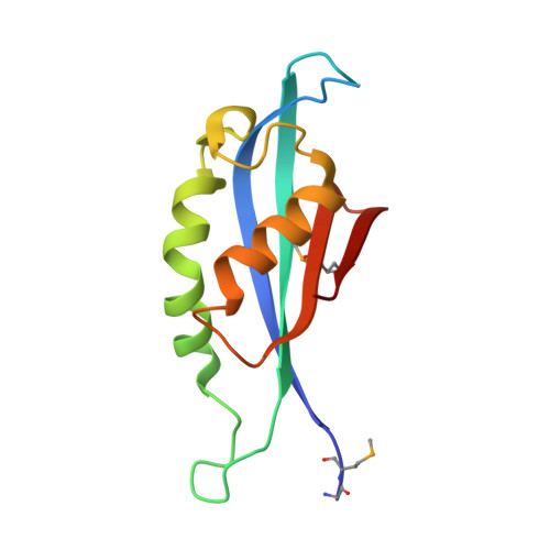
4NTM

QueD soaked with sepiapterin (selenomethionine substituted protein)


Experimental Data Snapshot

  • Method: X-RAY DIFFRACTION
  • Resolution: 2.05 Å
  • R-Value Free: 0.255 
  • R-Value Work: 0.225 
  • R-Value Observed: 0.226 

wwPDB Validation   3D Report Full Report


Ligand Structure Quality Assessment 


This is version 1.3 of the entry. See complete history


Literature

Biochemical and Structural Studies of 6-Carboxy-5,6,7,8-tetrahydropterin Synthase Reveal the Molecular Basis of Catalytic Promiscuity within the Tunnel-fold Superfamily.

Miles, Z.D.Roberts, S.A.McCarty, R.M.Bandarian, V.

(2014) J Biol Chem 289: 23641-23652

  • DOI: https://doi.org/10.1074/jbc.M114.555680
  • Primary Citation of Related Structures:  
    4NTK, 4NTM, 4NTN

  • PubMed Abstract: 

    6-Pyruvoyltetrahydropterin synthase (PTPS) homologs in both mammals and bacteria catalyze distinct reactions using the same 7,8-dihydroneopterin triphosphate substrate. The mammalian enzyme converts 7,8-dihydroneopterin triphosphate to 6-pyruvoyltetrahydropterin, whereas the bacterial enzyme catalyzes the formation of 6-carboxy-5,6,7,8-tetrahydropterin. To understand the basis for the differential activities we determined the crystal structure of a bacterial PTPS homolog in the presence and absence of various ligands. Comparison to mammalian structures revealed that although the active sites are nearly structurally identical, the bacterial enzyme houses a His/Asp dyad that is absent from the mammalian protein. Steady state and time-resolved kinetic analysis of the reaction catalyzed by the bacterial homolog revealed that these residues are responsible for the catalytic divergence. This study demonstrates how small variations in the active site can lead to the emergence of new functions in existing protein folds.


  • Organizational Affiliation

    From the Department of Chemistry and Biochemistry, University of Arizona, Tucson, Arizona 85721.


Macromolecules
Find similar proteins by:  (by identity cutoff)  |  3D Structure
Entity ID: 1
MoleculeChains Sequence LengthOrganismDetailsImage
6-carboxy-5,6,7,8-tetrahydropterin synthase
A, B, C, D, E
A, B, C, D, E, F
121Escherichia coli K-12Mutation(s): 0 
Gene Names: b2765JW2735queDygcM
EC: 4.1.2.50
UniProt
Find proteins for P65870 (Escherichia coli (strain K12))
Explore P65870 
Go to UniProtKB:  P65870
Entity Groups  
Sequence Clusters30% Identity50% Identity70% Identity90% Identity95% Identity100% Identity
UniProt GroupP65870
Sequence Annotations
Expand
  • Reference Sequence
Small Molecules
Ligands 2 Unique
IDChains Name / Formula / InChI Key2D Diagram3D Interactions
2K8
Query on 2K8

Download Ideal Coordinates CCD File 
H [auth A]
J [auth B]
L [auth C]
N [auth D]
P [auth E]
H [auth A],
J [auth B],
L [auth C],
N [auth D],
P [auth E],
R [auth F]
(6R)-2-amino-4-oxo-3,4,5,6,7,8-hexahydropteridine-6-carboxylic acid
C7 H9 N5 O3
QSIYONWVWDSRRO-UWTATZPHSA-N
ZN
Query on ZN

Download Ideal Coordinates CCD File 
G [auth A]
I [auth B]
K [auth C]
M [auth D]
O [auth E]
G [auth A],
I [auth B],
K [auth C],
M [auth D],
O [auth E],
Q [auth F]
ZINC ION
Zn
PTFCDOFLOPIGGS-UHFFFAOYSA-N
Modified Residues  1 Unique
IDChains TypeFormula2D DiagramParent
MSE
Query on MSE
A, B, C, D, E
A, B, C, D, E, F
L-PEPTIDE LINKINGC5 H11 N O2 SeMET
Experimental Data & Validation

Experimental Data

  • Method: X-RAY DIFFRACTION
  • Resolution: 2.05 Å
  • R-Value Free: 0.255 
  • R-Value Work: 0.225 
  • R-Value Observed: 0.226 
  • Space Group: P 41 21 2
Unit Cell:
Length ( Å )Angle ( ˚ )
a = 111.297α = 90
b = 111.297β = 90
c = 126.047γ = 90
Software Package:
Software NamePurpose
Blu-Icedata collection
REFMACrefinement
XDSdata reduction
XDSdata scaling

Structure Validation

View Full Validation Report


Ligand Structure Quality Assessment 


Entry History 

Deposition Data

Revision History  (Full details and data files)

  • Version 1.0: 2014-07-16
    Type: Initial release
  • Version 1.1: 2014-09-10
    Changes: Database references
  • Version 1.2: 2017-11-22
    Changes: Refinement description
  • Version 1.3: 2018-01-24
    Changes: Structure summary