4NRE

The structure of human 15-lipoxygenase-2 with a substrate mimic


Experimental Data Snapshot

  • Method: X-RAY DIFFRACTION
  • Resolution: 2.63 Å
  • R-Value Free: 0.203 
  • R-Value Work: 0.163 
  • R-Value Observed: 0.165 

wwPDB Validation   3D Report Full Report


Ligand Structure Quality Assessment 


This is version 1.3 of the entry. See complete history


Literature

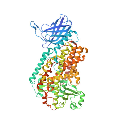
The structure of human 15-lipoxygenase-2 with a substrate mimic.

Kobe, M.J.Neau, D.B.Mitchell, C.E.Bartlett, S.G.Newcomer, M.E.

(2014) J Biol Chem 289: 8562-8569

  • DOI: https://doi.org/10.1074/jbc.M113.543777
  • Primary Citation of Related Structures:  
    4NRE

  • PubMed Abstract: 

    Atherosclerosis is associated with chronic inflammation occurring over decades. The enzyme 15-lipoxygenase-2 (15-LOX-2) is highly expressed in large atherosclerotic plaques, and its activity has been linked to the progression of macrophages to the lipid-laden foam cells present in atherosclerotic plaques. We report here the crystal structure of human 15-LOX-2 in complex with an inhibitor that appears to bind as a substrate mimic. 15-LOX-2 contains a long loop, composed of hydrophobic amino acids, which projects from the amino-terminal membrane-binding domain. The loop is flanked by two Ca(2+)-binding sites that confer Ca(2+)-dependent membrane binding. A comparison of the human 15-LOX-2 and 5-LOX structures reveals similarities at the active sites, as well striking differences that can be exploited for design of isoform-selective inhibitors.


  • Organizational Affiliation

    From the Department of Biological Sciences, Louisiana State University, Baton Rouge, Louisiana 70803 and.


Macromolecules
Find similar proteins by:  (by identity cutoff)  |  3D Structure
Entity ID: 1
MoleculeChains Sequence LengthOrganismDetailsImage
Arachidonate 15-lipoxygenase B696Homo sapiensMutation(s): 0 
Gene Names: ALOX15B
EC: 1.13.11.33 (PDB Primary Data), 1.13.11 (PDB Primary Data)
Membrane Entity: Yes 
UniProt & NIH Common Fund Data Resources
Find proteins for O15296 (Homo sapiens)
Explore O15296 
Go to UniProtKB:  O15296
PHAROS:  O15296
GTEx:  ENSG00000179593 
Entity Groups  
Sequence Clusters30% Identity50% Identity70% Identity90% Identity95% Identity100% Identity
UniProt GroupO15296
Sequence Annotations
Expand
  • Reference Sequence
Small Molecules
Ligands 5 Unique
IDChains Name / Formula / InChI Key2D Diagram3D Interactions
C8E
Query on C8E

Download Ideal Coordinates CCD File 
C [auth A],
D [auth A]
(HYDROXYETHYLOXY)TRI(ETHYLOXY)OCTANE
C16 H34 O5
FEOZZFHAVXYAMB-UHFFFAOYSA-N
SO4
Query on SO4

Download Ideal Coordinates CCD File 
E [auth A]
F [auth A]
G [auth A]
H [auth A]
I [auth A]
E [auth A],
F [auth A],
G [auth A],
H [auth A],
I [auth A],
J [auth A],
K [auth A],
L [auth A],
M [auth A],
N [auth A],
O [auth A],
P [auth A]
SULFATE ION
O4 S
QAOWNCQODCNURD-UHFFFAOYSA-L
GOL
Query on GOL

Download Ideal Coordinates CCD File 
S [auth A],
T [auth A],
U [auth A]
GLYCEROL
C3 H8 O3
PEDCQBHIVMGVHV-UHFFFAOYSA-N
FE2
Query on FE2

Download Ideal Coordinates CCD File 
B [auth A]FE (II) ION
Fe
CWYNVVGOOAEACU-UHFFFAOYSA-N
CA
Query on CA

Download Ideal Coordinates CCD File 
Q [auth A],
R [auth A]
CALCIUM ION
Ca
BHPQYMZQTOCNFJ-UHFFFAOYSA-N
Experimental Data & Validation

Experimental Data

  • Method: X-RAY DIFFRACTION
  • Resolution: 2.63 Å
  • R-Value Free: 0.203 
  • R-Value Work: 0.163 
  • R-Value Observed: 0.165 
  • Space Group: H 3 2
Unit Cell:
Length ( Å )Angle ( ˚ )
a = 155.794α = 90
b = 155.794β = 90
c = 263.219γ = 120
Software Package:
Software NamePurpose
XDSdata scaling
PHASERphasing
PHENIXrefinement
XDSdata reduction
SCALAdata scaling

Structure Validation

View Full Validation Report


Ligand Structure Quality Assessment 


Entry History 

Deposition Data

Revision History  (Full details and data files)

  • Version 1.0: 2014-02-12
    Type: Initial release
  • Version 1.1: 2014-02-26
    Changes: Other
  • Version 1.2: 2014-10-08
    Changes: Database references
  • Version 1.3: 2024-02-28
    Changes: Data collection, Database references, Derived calculations