Structural and energetic determinants of adhesive binding specificity in type I cadherins.
Vendome, J., Felsovalyi, K., Song, H., Yang, Z., Jin, X., Brasch, J., Harrison, O.J., Ahlsen, G., Bahna, F., Kaczynska, A., Katsamba, P.S., Edmond, D., Hubbell, W.L., Shapiro, L., Honig, B.(2014) Proc Natl Acad Sci U S A 111: E4175-E4184
- PubMed: 25253890 Search on PubMedSearch on PubMed Central
- DOI: https://doi.org/10.1073/pnas.1416737111
- Primary Citation Related Structures:
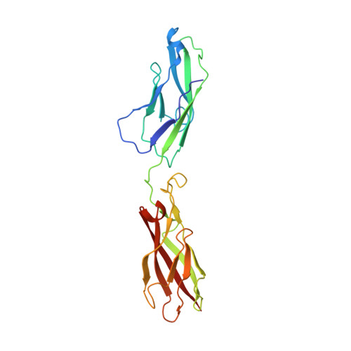
4NQQ, 4NUM, 4NUP, 4NUQ - PubMed Abstract:
Type I cadherin cell-adhesion proteins are similar in sequence and structure and yet are different enough to mediate highly specific cell-cell recognition phenomena. It has previously been shown that small differences in the homophilic and heterophilic binding affinities of different type I family members can account for the differential cell-sorting behavior. Here we use a combination of X-ray crystallography, analytical ultracentrifugation, surface plasmon resonance and double electron-electron resonance (DEER) electron paramagnetic resonance spectroscopy to identify the molecular determinants of type I cadherin dimerization affinities. Small changes in sequence are found to produce subtle structural and dynamical changes that impact relative affinities, in part via electrostatic and hydrophobic interactions, and in part through entropic effects because of increased conformational heterogeneity in the bound states as revealed by DEER distance mapping in the dimers. These findings highlight the remarkable ability of evolution to exploit a wide range of molecular properties to produce closely related members of the same protein family that have affinity differences finely tuned to mediate their biological roles.
- Department of Biochemistry and Molecular Biophysics, Center for Computational Biology and Bioinformatics, Department of Systems Biology, Howard Hughes Medical Institute, Columbia University, New York, NY 10032;
Organizational Affiliation: