4NQ2 | pdb_00004nq2

Structure of Zn(II)-bound metallo-beta-lactamse VIM-2 from Pseudomonas aeruginosa


Experimental Data Snapshot

  • Method: X-RAY DIFFRACTION
  • Resolution: 1.55 Å
  • R-Value Free: 
    0.209 (Depositor), 0.209 (DCC) 
  • R-Value Work: 
    0.176 (Depositor), 0.178 (DCC) 
  • R-Value Observed: 
    0.178 (Depositor) 

Starting Model: experimental
View more details

wwPDB Validation 3D Report Full Report

Validation slider image for 4NQ2

This is version 1.4 of the entry. See complete history

Literature

Biochemical, Mechanistic, and Spectroscopic Characterization of Metallo-beta-lactamase VIM-2.

Aitha, M.Marts, A.R.Bergstrom, A.Moller, A.J.Moritz, L.Turner, L.Nix, J.C.Bonomo, R.A.Page, R.C.Tierney, D.L.Crowder, M.W.

(2014) Biochemistry 53: 7321-7331

  • DOI: https://doi.org/10.1021/bi500916y
  • Primary Citation Related Structures: 
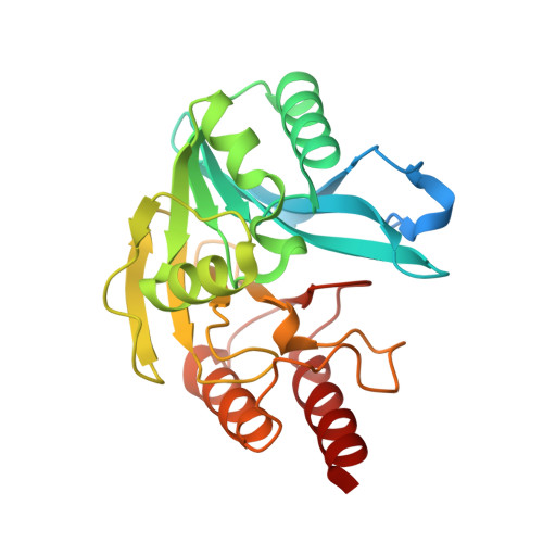
    4NQ2

  • PubMed Abstract: 

    This study examines metal binding to metallo-β-lactamase VIM-2, demonstrating the first successful preparation of a Co(II)-substituted VIM-2 analogue. Spectroscopic studies of the half- and fully metal loaded enzymes show that both Zn(II) and Co(II) bind cooperatively, where the major species present, regardless of stoichiometry, are apo- and di-Zn (or di-Co) enzymes. We determined the di-Zn VIM-2 structure to a resolution of 1.55 Å, and this structure supports results from spectroscopic studies. Kinetics, both steady-state and pre-steady-state, show that VIM-2 utilizes a mechanism that proceeds through a very short-lived anionic intermediate when chromacef is used as the substrate. Comparison with other B1 enzymes shows that those that bind Zn(II) cooperatively are better poised to protonate the intermediate on its formation, compared to those that bind Zn(II) non-cooperatively, which uniformly build up substantial amounts of the intermediate.


  • Organizational Affiliation
    • Department of Chemistry and Biochemistry, Miami University , 650 East High Street, Oxford, Ohio 45056, United States.

Macromolecule Content 

  • Total Structure Weight: 28.1 kDa 
  • Atom Count: 1,992 
  • Modeled Residue Count: 230 
  • Deposited Residue Count: 261 
  • Unique protein chains: 1

Macromolecules

Find similar proteins by:|  3D Structure
Entity ID: 1
MoleculeChains  Sequence LengthOrganismDetailsImage
Beta-lactamase class B VIM-2261Pseudomonas aeruginosaMutation(s): 0 
Gene Names: bla vim-2bla-VIM-2blasVIM-2blaVIM-2blaVIM2VIM-2
EC: 3.5.2.6
UniProt
Find proteins for Q9K2N0 (Pseudomonas aeruginosa)
Explore Q9K2N0 
Go to UniProtKB:  Q9K2N0
Entity Groups
Sequence Clusters30% Identity50% Identity70% Identity90% Identity95% Identity100% Identity
UniProt GroupQ9K2N0
Sequence Annotations
Expand
Reference Sequence

Experimental Data & Validation

Experimental Data

  • Method: X-RAY DIFFRACTION
  • Resolution: 1.55 Å
  • R-Value Free:  0.209 (Depositor), 0.209 (DCC) 
  • R-Value Work:  0.176 (Depositor), 0.178 (DCC) 
  • R-Value Observed: 0.178 (Depositor) 
Space Group: I 2 2 2
Unit Cell:
Length ( Å )Angle ( ˚ )
a = 68.294α = 90
b = 78.266β = 90
c = 79.767γ = 90
Software Package:
Software NamePurpose
JBluIce-EPICSdata collection
PHENIXmodel building
PHENIXrefinement
XDSdata reduction
SCALEPACKdata scaling
PHENIXphasing

Structure Validation

View Full Validation Report


Entry History 

Deposition Data

Revision History  (Full details and data files)

  • Version 1.0: 2014-11-12
    Type: Initial release
  • Version 1.1: 2014-12-10
    Changes: Database references
  • Version 1.2: 2017-08-09
    Changes: Database references
  • Version 1.3: 2017-11-22
    Changes: Refinement description
  • Version 1.4: 2023-09-20
    Changes: Data collection, Database references, Derived calculations, Refinement description