4NDR

Crystal structure Molybdenum Storage Protein with fully Mo-loaded cavity


Experimental Data Snapshot

  • Method: X-RAY DIFFRACTION
  • Resolution: 2.00 Å
  • R-Value Free: 0.191 
  • R-Value Work: 0.164 
  • R-Value Observed: 0.165 

wwPDB Validation   3D Report Full Report


Ligand Structure Quality Assessment 


This is version 1.4 of the entry. See complete history


Literature

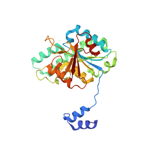
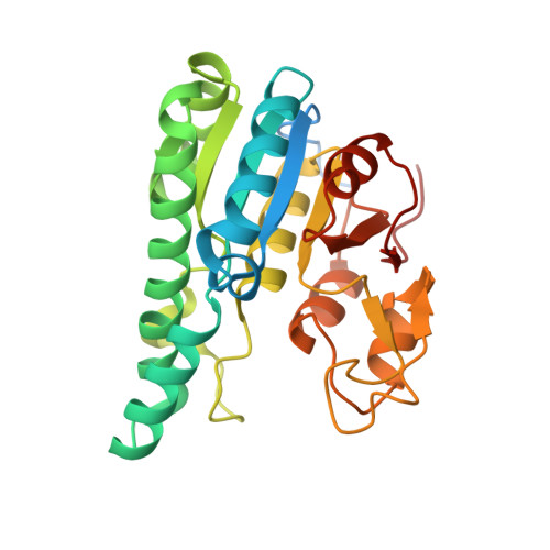
Structural diversity of polyoxomolybdate clusters along the three-fold axis of the molybdenum storage protein.

Poppe, J.Warkentin, E.Demmer, U.Kowalewski, B.Dierks, T.Schneider, K.Ermler, U.

(2014) J Inorg Biochem 138: 122-128

  • DOI: https://doi.org/10.1016/j.jinorgbio.2014.05.009
  • Primary Citation of Related Structures:  
    4NDO, 4NDP, 4NDQ, 4NDR

  • PubMed Abstract: 

    The molybdenum storage protein (MoSto) can store more than 100 Mo or W atoms as discrete polyoxometalate (POM) clusters. Here, we describe the three POM cluster sites along the threefold axis of the protein complex based on four X-ray structures with slightly different polyoxomolybdate compositions between 1.35 and 2 Å resolution. In contrast to the Moα-out binding site occupied by an Mo3 cluster, the Moα-in and Moβ binding sites contain rather weak and non-uniform electron density for the Mo atoms (but clearly identifiable by anomalous data), suggesting the presence of POM cluster ensembles and/or degradation products of larger aggregates. The "Moα-in cluster ensemble" was interpreted as an antiprism-like Mo6 species superimposed with an Mo7 pyramide and the "Moβ cluster ensemble" as an Mo13 cluster (present mostly in a degraded form) composed of a pyramidal Mo7 and a Mo3 building block linked by three spatially separated MoOx units. Inside the ball-shaped Mo13 cluster sits an occluded central atom, perhaps a metal ion. POM cluster formation at the Moα-in and Moβ sites appears to be driven by filtering out and binding/protecting self-assembled transient species complementary to the protein template.


  • Organizational Affiliation

    Max-Planck-Institut für Biophysik, Max-von-Laue-Straße 3, D-60438 Frankfurt am Main, Germany.


Macromolecules
Find similar proteins by:  (by identity cutoff)  |  3D Structure
Entity ID: 1
MoleculeChains Sequence LengthOrganismDetailsImage
Molybdenum storage protein subunit betaA [auth B]270Azotobacter vinelandii DJMutation(s): 0 
UniProt
Find proteins for P84253 (Azotobacter vinelandii (strain DJ / ATCC BAA-1303))
Explore P84253 
Go to UniProtKB:  P84253
Entity Groups  
Sequence Clusters30% Identity50% Identity70% Identity90% Identity95% Identity100% Identity
UniProt GroupP84253
Sequence Annotations
Expand
  • Reference Sequence
Find similar proteins by:  (by identity cutoff)  |  3D Structure
Entity ID: 2
MoleculeChains Sequence LengthOrganismDetailsImage
Molybdenum storage protein subunit alphaB [auth A]276Azotobacter vinelandii DJMutation(s): 0 
UniProt
Find proteins for P84308 (Azotobacter vinelandii (strain DJ / ATCC BAA-1303))
Explore P84308 
Go to UniProtKB:  P84308
Entity Groups  
Sequence Clusters30% Identity50% Identity70% Identity90% Identity95% Identity100% Identity
UniProt GroupP84308
Sequence Annotations
Expand
  • Reference Sequence
Small Molecules
Ligands 6 Unique
IDChains Name / Formula / InChI Key2D Diagram3D Interactions
8M0
Query on 8M0

Download Ideal Coordinates CCD File 
E [auth B],
X [auth A]
bis(mu4-oxo)-tetrakis(mu3-oxo)-hexakis(mu2-oxo)-hexadecaoxo-octamolybdenum (VI)
Mo8 O28
GSOSAILZTJNYOK-UHFFFAOYSA-N
ATP
Query on ATP

Download Ideal Coordinates CCD File 
C [auth B],
V [auth A]
ADENOSINE-5'-TRIPHOSPHATE
C10 H16 N5 O13 P3
ZKHQWZAMYRWXGA-KQYNXXCUSA-N
M10
Query on M10

Download Ideal Coordinates CCD File 
Y [auth A](mu3-oxo)-tris(mu2-oxo)-nonakisoxo-trimolybdenum (VI)
Mo3 O13
HGNGIYFURFJYFJ-UHFFFAOYSA-N
MO
Query on MO

Download Ideal Coordinates CCD File 
AA [auth A]
BA [auth A]
CA [auth A]
F [auth B]
G [auth B]
AA [auth A],
BA [auth A],
CA [auth A],
F [auth B],
G [auth B],
H [auth B],
I [auth B],
J [auth B],
K [auth B],
L [auth B],
M [auth B],
N [auth B],
O [auth B],
P [auth B],
Q [auth B],
R [auth B],
S [auth B],
T [auth B],
U [auth B],
Z [auth A]
MOLYBDENUM ATOM
Mo
ZOKXTWBITQBERF-UHFFFAOYSA-N
PO4
Query on PO4

Download Ideal Coordinates CCD File 
D [auth B]PHOSPHATE ION
O4 P
NBIIXXVUZAFLBC-UHFFFAOYSA-K
MG
Query on MG

Download Ideal Coordinates CCD File 
W [auth A]MAGNESIUM ION
Mg
JLVVSXFLKOJNIY-UHFFFAOYSA-N
Experimental Data & Validation

Experimental Data

  • Method: X-RAY DIFFRACTION
  • Resolution: 2.00 Å
  • R-Value Free: 0.191 
  • R-Value Work: 0.164 
  • R-Value Observed: 0.165 
  • Space Group: P 63 2 2
Unit Cell:
Length ( Å )Angle ( ˚ )
a = 114.27α = 90
b = 114.27β = 90
c = 234.14γ = 120
Software Package:
Software NamePurpose
PHENIXrefinement
REFMACrefinement
XDSdata reduction
XDSdata scaling
REFMACphasing

Structure Validation

View Full Validation Report


Ligand Structure Quality Assessment 


Entry History 

Deposition Data

Revision History  (Full details and data files)

  • Version 1.0: 2014-08-13
    Type: Initial release
  • Version 1.1: 2014-11-12
    Changes: Structure summary
  • Version 1.2: 2017-11-15
    Changes: Refinement description
  • Version 1.3: 2019-07-17
    Changes: Data collection, Refinement description
  • Version 1.4: 2023-09-20
    Changes: Data collection, Database references, Derived calculations, Refinement description