4N2X

Crystal Structure of DL-2-haloacid dehalogenase


Experimental Data Snapshot

  • Method: X-RAY DIFFRACTION
  • Resolution: 1.70 Å
  • R-Value Free: 0.172 
  • R-Value Work: 0.135 
  • R-Value Observed: 0.139 

wwPDB Validation   3D Report Full Report


This is version 1.1 of the entry. See complete history


Literature

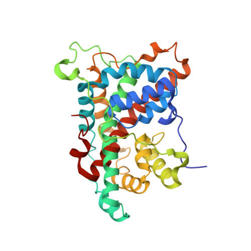
Binding modes of DL-2-haloacid dehalogenase revealed by crystallography, modeling and isotope effects studies.

Siwek, A.Omi, R.Hirotsu, K.Jitsumori, K.Esaki, N.Kurihara, T.Paneth, P.

(2013) Arch Biochem Biophys 540: 26-32

  • DOI: https://doi.org/10.1016/j.abb.2013.09.012
  • Primary Citation of Related Structures:  
    3WJ8, 4N2X

  • PubMed Abstract: 

    Several pathways of biotic dechlorination can be found in enzymes, each characterized by different chlorine isotopic fractionation, which can thus serve as a signature of a particular mechanism. Unlike other dehalogenases, DL-2-haloacid dehalogenase, DL-DEX, converts both enantiomers of the substrate. Chlorine isotope effects for this enzyme are larger than in the case of other dehalogenases. Recently, the 3D structure of this enzyme became available and enabled us to model these isotope effects and seek their origin. We show that the elevated values of the chlorine kinetic isotope effects originate in part in the processes of binding and migration within the enzyme active site that precede the dehalogenation step.


  • Organizational Affiliation

    Faculty of Pharmacy, Medical University of Lublin, Chodzki 4a, 20-093 Lublin, Poland; Faculty of Chemistry, Lodz University of Technology, Zeromskiego 116, 90-924 Lodz, Poland.


Macromolecules
Find similar proteins by:  (by identity cutoff)  |  3D Structure
Entity ID: 1
MoleculeChains Sequence LengthOrganismDetailsImage
DL-2-haloacid dehalogenase301Methylobacterium sp. CPA1Mutation(s): 0 
Gene Names: dl-dex
EC: 3.8.1.10
UniProt
Find proteins for A6BM74 (Methylobacterium sp. CPA1)
Explore A6BM74 
Go to UniProtKB:  A6BM74
Entity Groups  
Sequence Clusters30% Identity50% Identity70% Identity90% Identity95% Identity100% Identity
UniProt GroupA6BM74
Sequence Annotations
Expand
  • Reference Sequence
Small Molecules
Ligands 1 Unique
IDChains Name / Formula / InChI Key2D Diagram3D Interactions
GOL
Query on GOL

Download Ideal Coordinates CCD File 
G [auth A]
H [auth A]
I [auth B]
J [auth B]
K [auth D]
G [auth A],
H [auth A],
I [auth B],
J [auth B],
K [auth D],
L [auth D],
M [auth E],
N [auth E],
O [auth F],
P [auth F],
Q [auth G],
R [auth G]
GLYCEROL
C3 H8 O3
PEDCQBHIVMGVHV-UHFFFAOYSA-N
Experimental Data & Validation

Experimental Data

  • Method: X-RAY DIFFRACTION
  • Resolution: 1.70 Å
  • R-Value Free: 0.172 
  • R-Value Work: 0.135 
  • R-Value Observed: 0.139 
  • Space Group: P 63
Unit Cell:
Length ( Å )Angle ( ˚ )
a = 182.568α = 90
b = 182.568β = 90
c = 112.015γ = 120
Software Package:
Software NamePurpose
SCALEPACKdata scaling
REFMACrefinement
PDB_EXTRACTdata extraction
ADSCdata collection
HKL-2000data reduction

Structure Validation

View Full Validation Report


Entry History 

Deposition Data

Revision History  (Full details and data files)

  • Version 1.0: 2013-11-27
    Type: Initial release
  • Version 1.1: 2024-03-20
    Changes: Data collection, Database references, Derived calculations