4N1J | pdb_00004n1j

Crystal structures of NLRP14 pyrin domain reveal a conformational switch mechanism, regulating its molecular interactions


Experimental Data Snapshot

  • Method: X-RAY DIFFRACTION
  • Resolution: 2.60 Å
  • R-Value Free: 
    0.255 (Depositor), 0.265 (DCC) 
  • R-Value Work: 
    0.209 (Depositor), 0.225 (DCC) 
  • R-Value Observed: 
    0.212 (Depositor) 

wwPDB Validation 3D Report Full Report

Validation slider image for 4N1J

This is version 1.3 of the entry. See complete history

Literature

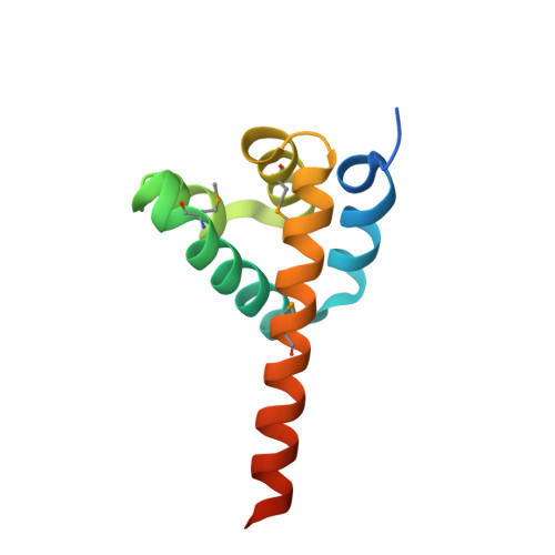
Structures of the NLRP14 pyrin domain reveal a conformational switch mechanism regulating its molecular interactions.

Eibl, C.Hessenberger, M.Wenger, J.Brandstetter, H.

(2014) Acta Crystallogr D Biol Crystallogr 70: 2007-2018

  • DOI: https://doi.org/10.1107/S1399004714010311
  • Primary Citation Related Structures: 
    4N1J, 4N1K, 4N1L

  • PubMed Abstract: 

    The cytosolic tripartite NLR receptors serve as important signalling platforms in innate immunity. While the C-terminal domains act as sensor and activation modules, the N-terminal death-like domain, e.g. the CARD or pyrin domain, is thought to recruit downstream effector molecules by homotypic interactions. Such homotypic complexes have been determined for all members of the death-domain superfamily except for pyrin domains. Here, crystal structures of human NLRP14 pyrin-domain variants are reported. The wild-type protein as well as the clinical D86V mutant reveal an unexpected rearrangement of the C-terminal helix α6, resulting in an extended α5/6 stem-helix. This reordering mediates a novel symmetric pyrin-domain dimerization mode. The conformational switching is controlled by a charge-relay system with a drastic impact on protein stability. How the identified charge relay allows classification of NLRP receptors with respect to distinct recruitment mechanisms is discussed.


  • Organizational Affiliation
    • Department of Molecular Biology, University of Salzburg, Billrothstrasse 11, 5020 Salzburg, Austria.

Macromolecule Content 

  • Total Structure Weight: 50.09 kDa 
  • Atom Count: 3,118 
  • Modeled Residue Count: 368 
  • Deposited Residue Count: 420 
  • Unique protein chains: 1

Macromolecules

Find similar proteins by:|  3D Structure
Entity ID: 1
MoleculeChains  Sequence LengthOrganismDetailsImage
NACHT, LRR and PYD domains-containing protein 14
A, B, C, D
105Homo sapiensMutation(s): 0 
Gene Names: NLRP14NALP14NOD5
UniProt & NIH Common Fund Data Resources
Find proteins for Q86W24 (Homo sapiens)
Explore Q86W24 
Go to UniProtKB:  Q86W24
PHAROS:  Q86W24
GTEx:  ENSG00000158077 
Entity Groups
Sequence Clusters30% Identity50% Identity70% Identity90% Identity95% Identity100% Identity
UniProt GroupQ86W24
Sequence Annotations
Expand
Reference Sequence

Experimental Data & Validation

Experimental Data

  • Method: X-RAY DIFFRACTION
  • Resolution: 2.60 Å
  • R-Value Free:  0.255 (Depositor), 0.265 (DCC) 
  • R-Value Work:  0.209 (Depositor), 0.225 (DCC) 
  • R-Value Observed: 0.212 (Depositor) 
Space Group: P 63
Unit Cell:
Length ( Å )Angle ( ˚ )
a = 89.61α = 90
b = 89.61β = 90
c = 107.871γ = 120
Software Package:
Software NamePurpose
MAR345data collection
Auto-Rickshawphasing
PHENIXrefinement
MOSFLMdata reduction
SCALAdata scaling

Structure Validation

View Full Validation Report


Entry History 

Deposition Data

Revision History  (Full details and data files)

  • Version 1.0: 2014-07-16
    Type: Initial release
  • Version 1.1: 2014-09-24
    Changes: Database references
  • Version 1.2: 2017-11-15
    Changes: Refinement description
  • Version 1.3: 2024-11-06
    Changes: Data collection, Database references, Derived calculations, Structure summary