4N1A

Thermomonospora curvata EccC (ATPases 2 and 3) in complex with a signal sequence peptide


Experimental Data Snapshot

  • Method: X-RAY DIFFRACTION
  • Resolution: 3.24 Å
  • R-Value Free: 0.266 
  • R-Value Work: 0.226 

wwPDB Validation   3D Report Full Report


Ligand Structure Quality Assessment 


This is version 1.2 of the entry. See complete history


Literature

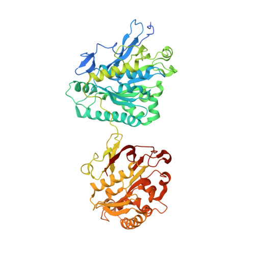
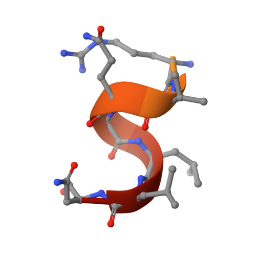
Substrates Control Multimerization and Activation of the Multi-Domain ATPase Motor of Type VII Secretion.

Rosenberg, O.S.Dovala, D.Li, X.Connolly, L.Bendebury, A.Finer-Moore, J.Holton, J.Cheng, Y.Stroud, R.M.Cox, J.S.

(2015) Cell 161: 501-512

  • DOI: https://doi.org/10.1016/j.cell.2015.03.040
  • Primary Citation of Related Structures:  
    4LWS, 4LYA, 4N1A, 4NH0

  • PubMed Abstract: 

    Mycobacterium tuberculosis and Staphylococcus aureus secrete virulence factors via type VII protein secretion (T7S), a system that intriguingly requires all of its secretion substrates for activity. To gain insights into T7S function, we used structural approaches to guide studies of the putative translocase EccC, a unique enzyme with three ATPase domains, and its secretion substrate EsxB. The crystal structure of EccC revealed that the ATPase domains are joined by linker/pocket interactions that modulate its enzymatic activity. EsxB binds via its signal sequence to an empty pocket on the C-terminal ATPase domain, which is accompanied by an increase in ATPase activity. Surprisingly, substrate binding does not activate EccC allosterically but, rather, by stimulating its multimerization. Thus, the EsxB substrate is also an integral T7S component, illuminating a mechanism that helps to explain interdependence of substrates, and suggests a model in which binding of substrates modulates their coordinate release from the bacterium.


  • Organizational Affiliation

    Division of Infectious Diseases, Department of Medicine, UCSF Medical Center, University of California, San Francisco, San Francisco, CA 94143-0654, USA.


Macromolecules
Find similar proteins by:  (by identity cutoff)  |  3D Structure
Entity ID: 1
MoleculeChains Sequence LengthOrganismDetailsImage
Cell divisionFtsK/SpoIIIEA,
B,
C,
G [auth E]
589Thermomonospora curvata DSM 43183Mutation(s): 0 
Gene Names: Tcur_0607
UniProt
Find proteins for D1A4G7 (Thermomonospora curvata (strain ATCC 19995 / DSM 43183 / JCM 3096 / KCTC 9072 / NBRC 15933 / NCIMB 10081 / Henssen B9))
Explore D1A4G7 
Go to UniProtKB:  D1A4G7
Entity Groups  
Sequence Clusters30% Identity50% Identity70% Identity90% Identity95% Identity100% Identity
UniProt GroupD1A4G7
Sequence Annotations
Expand
  • Reference Sequence

Find similar proteins by:  Sequence   |   3D Structure  

Entity ID: 2
MoleculeChains Sequence LengthOrganismDetailsImage
Uncharacterized proteinD [auth G],
E [auth H],
F [auth J],
H [auth K]
23Thermomonospora curvata DSM 43183Mutation(s): 0 
UniProt
Find proteins for D1A4H0 (Thermomonospora curvata (strain ATCC 19995 / DSM 43183 / JCM 3096 / KCTC 9072 / NBRC 15933 / NCIMB 10081 / Henssen B9))
Explore D1A4H0 
Go to UniProtKB:  D1A4H0
Entity Groups  
Sequence Clusters30% Identity50% Identity70% Identity90% Identity95% Identity100% Identity
UniProt GroupD1A4H0
Sequence Annotations
Expand
  • Reference Sequence
Small Molecules
Ligands 2 Unique
IDChains Name / Formula / InChI Key2D Diagram3D Interactions
ATP
Query on ATP

Download Ideal Coordinates CCD File 
I [auth A]
J [auth A]
M [auth B]
N [auth B]
Q [auth C]
I [auth A],
J [auth A],
M [auth B],
N [auth B],
Q [auth C],
R [auth C]
ADENOSINE-5'-TRIPHOSPHATE
C10 H16 N5 O13 P3
ZKHQWZAMYRWXGA-KQYNXXCUSA-N
MG
Query on MG

Download Ideal Coordinates CCD File 
K [auth A]
L [auth A]
O [auth B]
P [auth B]
S [auth C]
K [auth A],
L [auth A],
O [auth B],
P [auth B],
S [auth C],
T [auth C],
U [auth E],
V [auth E]
MAGNESIUM ION
Mg
JLVVSXFLKOJNIY-UHFFFAOYSA-N
Experimental Data & Validation

Experimental Data

  • Method: X-RAY DIFFRACTION
  • Resolution: 3.24 Å
  • R-Value Free: 0.266 
  • R-Value Work: 0.226 
  • Space Group: P 41 21 2
Unit Cell:
Length ( Å )Angle ( ˚ )
a = 216.15α = 90
b = 216.15β = 90
c = 186.51γ = 90
Software Package:
Software NamePurpose
HKL-2000data collection
PHASERphasing
PHENIXrefinement
HKL-2000data reduction
HKL-2000data scaling

Structure Validation

View Full Validation Report


Ligand Structure Quality Assessment 


Entry History 

Deposition Data

Revision History  (Full details and data files)

  • Version 1.0: 2015-02-11
    Type: Initial release
  • Version 1.1: 2016-09-21
    Changes: Database references
  • Version 1.2: 2024-02-28
    Changes: Data collection, Database references, Derived calculations