4MW4

Trypanosoma brucei methionyl-tRNA synthetase in complex with inhibitor 1-{3-[(5-chloro-2-hydroxy-3-iodobenzyl)amino]propyl}-3-thiophen-3-ylurea (Chem 1473)


Experimental Data Snapshot

  • Method: X-RAY DIFFRACTION
  • Resolution: 2.50 Å
  • R-Value Free: 0.231 
  • R-Value Work: 0.204 
  • R-Value Observed: 0.206 

wwPDB Validation   3D Report Full Report


Ligand Structure Quality Assessment 


This is version 1.1 of the entry. See complete history


Literature

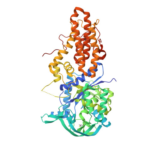
Structures of Trypanosoma brucei Methionyl-tRNA Synthetase with Urea-Based Inhibitors Provide Guidance for Drug Design against Sleeping Sickness.

Koh, C.Y.Kim, J.E.Wetzel, A.B.de van der Schueren, W.J.Shibata, S.Ranade, R.M.Liu, J.Zhang, Z.Gillespie, J.R.Buckner, F.S.Verlinde, C.L.Fan, E.Hol, W.G.

(2014) PLoS Negl Trop Dis 8: e2775-e2775

  • DOI: https://doi.org/10.1371/journal.pntd.0002775
  • Primary Citation of Related Structures:  
    4MVW, 4MVX, 4MVY, 4MW0, 4MW1, 4MW2, 4MW4, 4MW5, 4MW6, 4MW7, 4MW9, 4MWB, 4MWC, 4MWD, 4MWE

  • PubMed Abstract: 

    Methionyl-tRNA synthetase of Trypanosoma brucei (TbMetRS) is an important target in the development of new antitrypanosomal drugs. The enzyme is essential, highly flexible and displaying a large degree of changes in protein domains and binding pockets in the presence of substrate, product and inhibitors. Targeting this protein will benefit from a profound understanding of how its structure adapts to ligand binding. A series of urea-based inhibitors (UBIs) has been developed with IC50 values as low as 19 nM against the enzyme. The UBIs were shown to be orally available and permeable through the blood-brain barrier, and are therefore candidates for development of drugs for the treatment of late stage human African trypanosomiasis. Here, we expand the structural diversity of inhibitors from the previously reported collection and tested for their inhibitory effect on TbMetRS and on the growth of T. brucei cells. The binding modes and binding pockets of 14 UBIs are revealed by determination of their crystal structures in complex with TbMetRS at resolutions between 2.2 Å to 2.9 Å. The structures show binding of the UBIs through conformational selection, including occupancy of the enlarged methionine pocket and the auxiliary pocket. General principles underlying the affinity of UBIs for TbMetRS are derived from these structures, in particular the optimum way to fill the two binding pockets. The conserved auxiliary pocket might play a role in binding tRNA. In addition, a crystal structure of a ternary TbMetRS•inhibitor•AMPPCP complex indicates that the UBIs are not competing with ATP for binding, instead are interacting with ATP through hydrogen bond. This suggests a possibility that a general 'ATP-engaging' binding mode can be utilized for the design and development of inhibitors targeting tRNA synthetases of other disease-causing pathogen.


  • Organizational Affiliation

    Department of Biochemistry, University of Washington, Seattle, Washington, United States of America.


Macromolecules
Find similar proteins by:  (by identity cutoff)  |  3D Structure
Entity ID: 1
MoleculeChains Sequence LengthOrganismDetailsImage
Methionyl-tRNA synthetase
A, B
542Trypanosoma bruceiMutation(s): 3 
Gene Names: Tb10.70.6470
EC: 6.1.1.10
UniProt
Find proteins for Q38C91 (Trypanosoma brucei brucei (strain 927/4 GUTat10.1))
Explore Q38C91 
Go to UniProtKB:  Q38C91
Entity Groups  
Sequence Clusters30% Identity50% Identity70% Identity90% Identity95% Identity100% Identity
UniProt GroupQ38C91
Sequence Annotations
Expand
  • Reference Sequence
Small Molecules
Ligands 5 Unique
IDChains Name / Formula / InChI Key2D Diagram3D Interactions
2EB
Query on 2EB

Download Ideal Coordinates CCD File 
U [auth B]1-{3-[(5-chloro-2-hydroxy-3-iodobenzyl)amino]propyl}-3-thiophen-3-ylurea
C15 H17 Cl I N3 O2 S
UJOHHPWMXXTHSR-UHFFFAOYSA-N
MET
Query on MET

Download Ideal Coordinates CCD File 
K [auth A]METHIONINE
C5 H11 N O2 S
FFEARJCKVFRZRR-BYPYZUCNSA-N
SO4
Query on SO4

Download Ideal Coordinates CCD File 
J [auth A],
S [auth B],
T [auth B]
SULFATE ION
O4 S
QAOWNCQODCNURD-UHFFFAOYSA-L
GOL
Query on GOL

Download Ideal Coordinates CCD File 
C [auth A]
D [auth A]
E [auth A]
F [auth A]
L [auth B]
C [auth A],
D [auth A],
E [auth A],
F [auth A],
L [auth B],
M [auth B],
N [auth B],
O [auth B],
P [auth B]
GLYCEROL
C3 H8 O3
PEDCQBHIVMGVHV-UHFFFAOYSA-N
DMS
Query on DMS

Download Ideal Coordinates CCD File 
G [auth A],
H [auth A],
I [auth A],
Q [auth B],
R [auth B]
DIMETHYL SULFOXIDE
C2 H6 O S
IAZDPXIOMUYVGZ-UHFFFAOYSA-N
Modified Residues  1 Unique
IDChains TypeFormula2D DiagramParent
CAS
Query on CAS
A, B
L-PEPTIDE LINKINGC5 H12 As N O2 SCYS
Experimental Data & Validation

Experimental Data

  • Method: X-RAY DIFFRACTION
  • Resolution: 2.50 Å
  • R-Value Free: 0.231 
  • R-Value Work: 0.204 
  • R-Value Observed: 0.206 
  • Space Group: P 21 21 21
Unit Cell:
Length ( Å )Angle ( ˚ )
a = 87.829α = 90
b = 106.063β = 90
c = 207.642γ = 90
Software Package:
Software NamePurpose
DENZOdata reduction
SCALEPACKdata scaling
PHASERphasing
REFMACrefinement
PDB_EXTRACTdata extraction
HKL-2000data collection

Structure Validation

View Full Validation Report


Ligand Structure Quality Assessment 


Entry History 

Deposition Data

Revision History  (Full details and data files)

  • Version 1.0: 2014-04-30
    Type: Initial release
  • Version 1.1: 2023-09-20
    Changes: Data collection, Database references, Derived calculations, Refinement description