Structural Basis for Autoinhibition of CTP:Phosphocholine Cytidylyltransferase (CCT), the Regulatory Enzyme in Phosphatidylcholine Synthesis, by Its Membrane-binding Amphipathic Helix.
Lee, J., Taneva, S.G., Holland, B.W., Tieleman, D.P., Cornell, R.B.(2014) J Biol Chem 289: 1742-1755
- PubMed: 24275660 Search on PubMedSearch on PubMed Central
- DOI: https://doi.org/10.1074/jbc.M113.526970
- Primary Citation Related Structures:
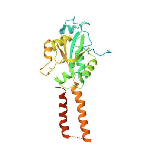
4MVC, 4MVD - PubMed Abstract:
CTP:phosphocholine cytidylyltransferase (CCT) interconverts between an inactive soluble and active membrane-bound form in response to changes in membrane lipid composition. Activation involves disruption of an inhibitory interaction between the αE helices at the base of the active site and an autoinhibitory (AI) segment in the regulatory M domain and membrane insertion of the M domain as an amphipathic helix. We show that in the CCT soluble form the AI segment functions to suppress kcat and elevate the Km for CTP. The crystal structure of a CCT dimer composed of the catalytic and AI segments reveals an AI-αE interaction as a cluster of four amphipathic helices (two αE and two AI helices) at the base of the active sites. This interaction corroborates mutagenesis implicating multiple hydrophobic residues within the AI segment that contribute to its silencing function. The AI-αE interaction directs the turn at the C-terminal end of the AI helix into backbone-to-backbone contact with a loop (L2) at the opening to the active site, which houses the key catalytic residue, lysine 122. Molecular dynamics simulations suggest that lysine 122 side-chain orientations are constrained by contacts with the AI helix-turn, which could obstruct its engagement with substrates. This work deciphers how the CCT regulatory amphipathic helix functions as a silencing device.
- From the Departments of Molecular Biology and Biochemistry and.
Organizational Affiliation: