4MQM | pdb_00004mqm

Crystal structure of Antigen 85C in presence of Ebselen


Experimental Data Snapshot

  • Method: X-RAY DIFFRACTION
  • Resolution: 1.35 Å
  • R-Value Free: 
    0.191 (Depositor), 0.189 (DCC) 
  • R-Value Work: 
    0.170 (Depositor), 0.168 (DCC) 
  • R-Value Observed: 
    0.171 (Depositor) 

wwPDB Validation 3D Report Full Report

Validation slider image for 4MQM

This is version 1.2 of the entry. See complete history

Literature

Mechanism of inhibition of Mycobacterium tuberculosis antigen 85 by ebselen.

Favrot, L.Grzegorzewicz, A.E.Lajiness, D.H.Marvin, R.K.Boucau, J.Isailovic, D.Jackson, M.Ronning, D.R.

(2013) Nat Commun 4: 2748-2748

  • DOI: https://doi.org/10.1038/ncomms3748
  • Primary Citation Related Structures: 
    4MQL, 4MQM

  • PubMed Abstract: 

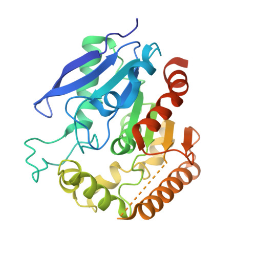
    The increasing prevalence of drug-resistant tuberculosis highlights the need for identifying new antitubercular drugs that can treat these infections. The antigen 85 (Ag85) complex has emerged as an intriguing mycobacterial drug target due to its central role in synthesizing major components of the inner and outer leaflets of the mycobacterial outer membrane. Here we identify ebselen (EBS) as a potent inhibitor of the Mycobacterium tuberculosis Ag85 complex. Mass spectrometry data show that EBS binds covalently to a cysteine residue (C209) located near the Ag85C active site. The crystal structure of Ag85C in the presence of EBS shows that C209 modification restructures the active site, thereby disrupting the hydrogen-bonded network within the active site that is essential for enzymatic activity. C209 mutations display marked decreases in enzymatic activity. These data suggest that compounds using this mechanism of action will strongly inhibit the Ag85 complex and minimize the selection of drug resistance.


  • Organizational Affiliation
    • Department of Chemistry, University of Toledo, Toledo, Ohio 43606-3390, USA.

Macromolecule Content 

  • Total Structure Weight: 33.31 kDa 
  • Atom Count: 2,268 
  • Modeled Residue Count: 269 
  • Deposited Residue Count: 304 
  • Unique protein chains: 1

Macromolecules

Find similar proteins by:|  3D Structure
Entity ID: 1
MoleculeChains  Sequence LengthOrganismDetailsImage
Diacylglycerol acyltransferase/mycolyltransferase Ag85C304Mycobacterium tuberculosisMutation(s): 0 
Gene Names: fbpCmpt45Rv0129cMT0137MTCI5.03c
EC: 2.3.1.122 (PDB Primary Data), 2.3.1.20 (PDB Primary Data)
UniProt
Find proteins for P9WQN9 (Mycobacterium tuberculosis (strain ATCC 25618 / H37Rv))
Explore P9WQN9 
Go to UniProtKB:  P9WQN9
Entity Groups
Sequence Clusters30% Identity50% Identity70% Identity90% Identity95% Identity100% Identity
UniProt GroupP9WQN9
Sequence Annotations
Expand
Reference Sequence

Experimental Data & Validation

Experimental Data

  • Method: X-RAY DIFFRACTION
  • Resolution: 1.35 Å
  • R-Value Free:  0.191 (Depositor), 0.189 (DCC) 
  • R-Value Work:  0.170 (Depositor), 0.168 (DCC) 
  • R-Value Observed: 0.171 (Depositor) 
Space Group: P 21 21 21
Unit Cell:
Length ( Å )Angle ( ˚ )
a = 60.643α = 90
b = 68.119β = 90
c = 75.104γ = 90
Software Package:
Software NamePurpose
HKL-2000data collection
PHENIXmodel building
PHENIXrefinement
HKL-2000data reduction
HKL-2000data scaling
PHENIXphasing

Structure Validation

View Full Validation Report


Entry History 

Deposition Data

Revision History  (Full details and data files)

  • Version 1.0: 2013-11-13
    Type: Initial release
  • Version 1.1: 2013-12-25
    Changes: Database references
  • Version 1.2: 2024-02-28
    Changes: Data collection, Database references