4MOC | pdb_00004moc

Human Acyl-coenzyme A Thioesterase 12


Experimental Data Snapshot

  • Method: X-RAY DIFFRACTION
  • Resolution: 2.50 Å
  • R-Value Free: 
    0.247 (Depositor), 0.250 (DCC) 
  • R-Value Work: 
    0.202 (Depositor), 0.203 (DCC) 
  • R-Value Observed: 
    0.204 (Depositor) 

wwPDB Validation 3D Report Full Report

Validation slider image for 4MOC

Ligand Structure Quality Assessment 


This is version 1.5 of the entry. See complete history

Literature

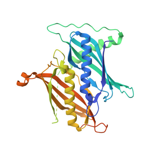
Structural basis for regulation of the human acetyl-CoA thioesterase 12 and interactions with the steroidogenic acute regulatory protein-related lipid transfer (START) domain.

Swarbrick, C.M.Roman, N.Cowieson, N.Patterson, E.I.Nanson, J.Siponen, M.I.Berglund, H.Lehtio, L.Forwood, J.K.

(2014) J Biological Chem 289: 24263-24274

  • DOI: https://doi.org/10.1074/jbc.M114.589408
  • Primary Citation Related Structures: 
    4MOB, 4MOC

  • PubMed Abstract: 

    Acetyl-CoA plays a fundamental role in cell signaling and metabolic pathways, with its cellular levels tightly controlled through reciprocal regulation of enzymes that mediate its synthesis and catabolism. ACOT12, the primary acetyl-CoA thioesterase in the liver of human, mouse, and rat, is responsible for cleavage of the thioester bond within acetyl-CoA, producing acetate and coenzyme A for a range of cellular processes. The enzyme is regulated by ADP and ATP, which is believed to be mediated through the ligand-induced oligomerization of the thioesterase domains, whereby ATP induces active dimers and tetramers, whereas apo- and ADP-bound ACOT12 are monomeric and inactive. Here, using a range of structural and biophysical techniques, it is demonstrated that ACOT12 is a trimer rather than a tetramer and that neither ADP nor ATP exert their regulatory effects by altering the oligomeric status of the enzyme. Rather, the binding site and mechanism of ADP regulation have been determined to occur through two novel regulatory regions, one involving a large loop that links the thioesterase domains (Phe(154)-Thr(178)), defined here as RegLoop1, and a second region involving the C terminus of thioesterase domain 2 (Gln(304)-Gly(326)), designated RegLoop2. Mutagenesis confirmed that Arg(312) and Arg(313) are crucial for this mode of regulation, and novel interactions with the START domain are presented together with insights into domain swapping within eukaryotic thioesterases for substrate recognition. In summary, these experiments provide the first structural insights into the regulation of this enzyme family, revealing an alternate hypothesis likely to be conserved throughout evolution.


  • Organizational Affiliation
    • From the School of Biomedical Sciences, Charles Sturt University, Wagga Wagga, New South Wales 2678, Australia.

Macromolecule Content 

  • Total Structure Weight: 37.88 kDa 
  • Atom Count: 2,240 
  • Modeled Residue Count: 273 
  • Deposited Residue Count: 332 
  • Unique protein chains: 1

Macromolecules

Find similar proteins by:|  3D Structure
Entity ID: 1
MoleculeChains  Sequence LengthOrganismDetailsImage
Acyl-coenzyme A thioesterase 12332Homo sapiensMutation(s): 0 
Gene Names: ACOT12CACHCACH1STARD15
EC: 3.1.2.1
UniProt & NIH Common Fund Data Resources
Find proteins for Q8WYK0 (Homo sapiens)
Explore Q8WYK0 
Go to UniProtKB:  Q8WYK0
PHAROS:  Q8WYK0
GTEx:  ENSG00000172497 
Entity Groups
Sequence Clusters30% Identity50% Identity70% Identity90% Identity95% Identity100% Identity
UniProt GroupQ8WYK0
Sequence Annotations
Expand
Reference Sequence

Small Molecules

Ligands 1 Unique
IDChains Name / Formula / InChI Key2D Diagram3D Interactions
COA

Query on COA


Download:Ideal Coordinates CCD File
B [auth A]COENZYME A
C21 H36 N7 O16 P3 S
RGJOEKWQDUBAIZ-IBOSZNHHSA-N

Experimental Data & Validation

Experimental Data

  • Method: X-RAY DIFFRACTION
  • Resolution: 2.50 Å
  • R-Value Free:  0.247 (Depositor), 0.250 (DCC) 
  • R-Value Work:  0.202 (Depositor), 0.203 (DCC) 
  • R-Value Observed: 0.204 (Depositor) 
Space Group: P 3 2 1
Unit Cell:
Length ( Å )Angle ( ˚ )
a = 123.56α = 90
b = 123.56β = 90
c = 49.75γ = 120
Software Package:
Software NamePurpose
Blu-Icedata collection
PHASERphasing
PHENIXrefinement
MOSFLMdata reduction
Aimlessdata scaling

Structure Validation

View Full Validation Report


Ligand Structure Quality Assessment 


Entry History 

Deposition Data

Revision History  (Full details and data files)

  • Version 1.0: 2014-07-16
    Type: Initial release
  • Version 1.1: 2014-07-23
    Changes: Database references
  • Version 1.2: 2015-04-01
    Changes: Database references
  • Version 1.3: 2017-11-15
    Changes: Refinement description
  • Version 1.4: 2018-01-24
    Changes: Structure summary
  • Version 1.5: 2024-02-28
    Changes: Data collection, Database references, Derived calculations