4MNG

Structure of the DP10.7 TCR with CD1d-sulfatide


Experimental Data Snapshot

  • Method: X-RAY DIFFRACTION
  • Resolution: 3.01 Å
  • R-Value Free: 0.207 
  • R-Value Work: 0.178 
  • R-Value Observed: 0.179 

wwPDB Validation   3D Report Full Report


Ligand Structure Quality Assessment 


This is version 1.4 of the entry. See complete history


Literature

Crystal Structure of V delta 1 T Cell Receptor in Complex with CD1d-Sulfatide Shows MHC-like Recognition of a Self-Lipid by Human gamma delta T Cells.

Luoma, A.M.Castro, C.D.Mayassi, T.Bembinster, L.A.Bai, L.Picard, D.Anderson, B.Scharf, L.Kung, J.E.Sibener, L.V.Savage, P.B.Jabri, B.Bendelac, A.Adams, E.J.

(2013) Immunity 39: 1032-1042

  • DOI: https://doi.org/10.1016/j.immuni.2013.11.001
  • Primary Citation of Related Structures:  
    4MNG, 4MNH, 4MQ7, 4NDM

  • PubMed Abstract: 

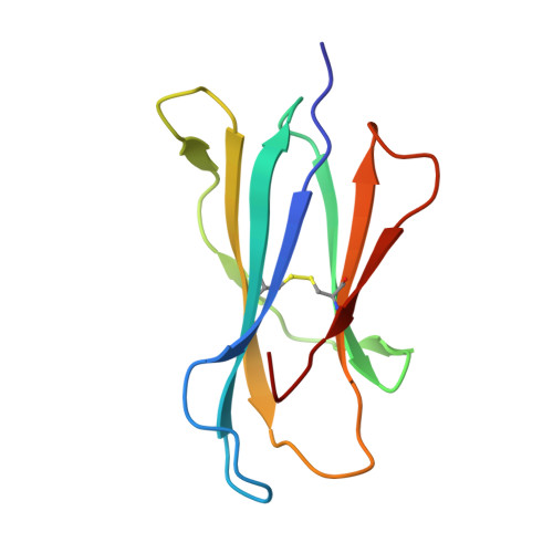
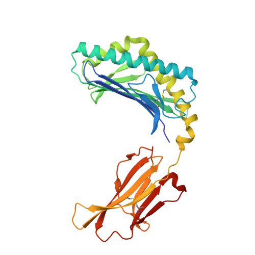
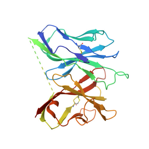
    The nature of the antigens recognized by γδ T cells and their potential recognition of major histocompatibility complex (MHC)-like molecules has remained unclear. Members of the CD1 family of lipid-presenting molecules are suggested ligands for Vδ1 TCR-expressing γδ T cells, the major γδ lymphocyte population in epithelial tissues. We crystallized a Vδ1 TCR in complex with CD1d and the self-lipid sulfatide, revealing the unusual recognition of CD1d by germline Vδ1 residues spanning all complementarity-determining region (CDR) loops, as well as sulfatide recognition separately encoded by nongermline CDR3δ residues. Binding and functional analysis showed that CD1d presenting self-lipids, including sulfatide, was widely recognized by gut Vδ1+ γδ T cells. These findings provide structural demonstration of MHC-like recognition of a self-lipid by γδ T cells and reveal the prevalence of lipid recognition by innate-like T cell populations.


  • Organizational Affiliation

    Committee on Immunology, University of Chicago, Chicago, IL 60637, USA; Department of Biochemistry and Molecular Biology, University of Chicago, Chicago, IL 60637, USA.


Macromolecules
Find similar proteins by:  (by identity cutoff)  |  3D Structure
Entity ID: 1
MoleculeChains Sequence LengthOrganismDetailsImage
Beta-2-microglobulinA [auth B],
C [auth D]
99Mus musculusMutation(s): 0 
Gene Names: B2m
UniProt & NIH Common Fund Data Resources
Find proteins for P01887 (Mus musculus)
Explore P01887 
Go to UniProtKB:  P01887
IMPC:  MGI:88127
Entity Groups  
Sequence Clusters30% Identity50% Identity70% Identity90% Identity95% Identity100% Identity
UniProt GroupP01887
Sequence Annotations
Expand
  • Reference Sequence
Find similar proteins by:  (by identity cutoff)  |  3D Structure
Entity ID: 2
MoleculeChains Sequence LengthOrganismDetailsImage
Cd1d1 proteinB [auth A],
D [auth C]
281Mus musculusMutation(s): 0 
UniProt & NIH Common Fund Data Resources
Find proteins for P11609 (Mus musculus)
Explore P11609 
Go to UniProtKB:  P11609
IMPC:  MGI:107674
Find proteins for P15813 (Homo sapiens)
Explore P15813 
Go to UniProtKB:  P15813
PHAROS:  P15813
GTEx:  ENSG00000158473 
Entity Groups  
Sequence Clusters30% Identity50% Identity70% Identity90% Identity95% Identity100% Identity
UniProt GroupsP11609P15813
Sequence Annotations
Expand
  • Reference Sequence
Find similar proteins by:  (by identity cutoff)  |  3D Structure
Entity ID: 3
MoleculeChains Sequence LengthOrganismDetailsImage
TRA@ protein,TRA@ protein, Ti antigen CD3-associated protein gamma chain V-J-C region
E, F
262Homo sapiensMutation(s): 0 
Gene Names: TRA@
UniProt
Find proteins for Q6PJ56 (Homo sapiens)
Explore Q6PJ56 
Go to UniProtKB:  Q6PJ56
Entity Groups  
Sequence Clusters30% Identity50% Identity70% Identity90% Identity95% Identity100% Identity
UniProt GroupQ6PJ56
Sequence Annotations
Expand
  • Reference Sequence
Small Molecules
Ligands 2 Unique
IDChains Name / Formula / InChI Key2D Diagram3D Interactions
CIS
Query on CIS

Download Ideal Coordinates CCD File 
J [auth A],
N [auth C]
(15Z)-N-((1S,2R,3E)-2-HYDROXY-1-{[(3-O-SULFO-BETA-D-GALACTOPYRANOSYL)OXY]METHYL}HEPTADEC-3-ENYL)TETRACOS-15-ENAMIDE
C48 H91 N O11 S
ZZQWQNAZXFNSEP-JCOQVFCVSA-N
NAG
Query on NAG

Download Ideal Coordinates CCD File 
G [auth A]
H [auth A]
I [auth A]
K [auth C]
L [auth C]
G [auth A],
H [auth A],
I [auth A],
K [auth C],
L [auth C],
M [auth C]
2-acetamido-2-deoxy-beta-D-glucopyranose
C8 H15 N O6
OVRNDRQMDRJTHS-FMDGEEDCSA-N
Experimental Data & Validation

Experimental Data

  • Method: X-RAY DIFFRACTION
  • Resolution: 3.01 Å
  • R-Value Free: 0.207 
  • R-Value Work: 0.178 
  • R-Value Observed: 0.179 
  • Space Group: P 32
Unit Cell:
Length ( Å )Angle ( ˚ )
a = 229.333α = 90
b = 229.333β = 90
c = 52.646γ = 120
Software Package:
Software NamePurpose
HKL-2000data collection
PHASERphasing
PHENIXrefinement
HKL-2000data reduction
HKL-2000data scaling

Structure Validation

View Full Validation Report


Ligand Structure Quality Assessment 


Entry History 

Deposition Data

Revision History  (Full details and data files)

  • Version 1.0: 2013-12-18
    Type: Initial release
  • Version 1.1: 2014-01-01
    Changes: Database references
  • Version 1.2: 2017-06-14
    Changes: Database references, Source and taxonomy, Structure summary
  • Version 1.3: 2020-07-29
    Type: Remediation
    Reason: Carbohydrate remediation
    Changes: Data collection, Derived calculations, Structure summary
  • Version 1.4: 2023-09-20
    Changes: Data collection, Database references, Refinement description, Structure summary