4MNE

Crystal structure of the BRAF:MEK1 complex


Experimental Data Snapshot

  • Method: X-RAY DIFFRACTION
  • Resolution: 2.85 Å
  • R-Value Free: 0.240 
  • R-Value Work: 0.185 
  • R-Value Observed: 0.186 

wwPDB Validation   3D Report Full Report


Ligand Structure Quality Assessment 


This is version 1.3 of the entry. See complete history


Literature

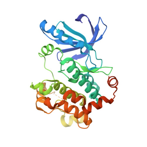
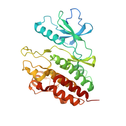
Structure of the BRAF-MEK Complex Reveals a Kinase Activity Independent Role for BRAF in MAPK Signaling.

Haling, J.R.Sudhamsu, J.Yen, I.Sideris, S.Sandoval, W.Phung, W.Bravo, B.J.Giannetti, A.M.Peck, A.Masselot, A.Morales, T.Smith, D.Brandhuber, B.J.Hymowitz, S.G.Malek, S.

(2014) Cancer Cell 26: 402-413

  • DOI: https://doi.org/10.1016/j.ccr.2014.07.007
  • Primary Citation of Related Structures:  
    4MNE, 4MNF

  • PubMed Abstract: 

    Numerous oncogenic mutations occur within the BRAF kinase domain (BRAF(KD)). Here we show that stable BRAF-MEK1 complexes are enriched in BRAF(WT) and KRAS mutant (MT) cells but not in BRAF(MT) cells. The crystal structure of the BRAF(KD) in a complex with MEK1 reveals a face-to-face dimer sensitive to MEK1 phosphorylation but insensitive to BRAF dimerization. Structure-guided studies reveal that oncogenic BRAF mutations function by bypassing the requirement for BRAF dimerization for activity or weakening the interaction with MEK1. Finally, we show that conformation-specific BRAF inhibitors can sequester a dormant BRAF-MEK1 complex resulting in pathway inhibition. Taken together, these findings reveal a regulatory role for BRAF in the MAPK pathway independent of its kinase activity but dependent on interaction with MEK.


  • Organizational Affiliation

    Department of Biochemical and Cellular Pharmacology, Genentech Inc., 1 DNA Way, South San Francisco, CA 94080, USA.


Macromolecules
Find similar proteins by:  (by identity cutoff)  |  3D Structure
Entity ID: 1
MoleculeChains Sequence LengthOrganismDetailsImage
Dual specificity mitogen-activated protein kinase kinase 1
A, D, E, H
341Homo sapiensMutation(s): 0 
Gene Names: MAP2K1MEK1PRKMK1
EC: 2.7.12.2
UniProt & NIH Common Fund Data Resources
Find proteins for Q02750 (Homo sapiens)
Explore Q02750 
Go to UniProtKB:  Q02750
PHAROS:  Q02750
GTEx:  ENSG00000169032 
Entity Groups  
Sequence Clusters30% Identity50% Identity70% Identity90% Identity95% Identity100% Identity
UniProt GroupQ02750
Sequence Annotations
Expand
  • Reference Sequence
Find similar proteins by:  (by identity cutoff)  |  3D Structure
Entity ID: 2
MoleculeChains Sequence LengthOrganismDetailsImage
Serine/threonine-protein kinase B-raf
B, C, F, G
308Homo sapiensMutation(s): 0 
Gene Names: BRAFBRAF1RAFB1
EC: 2.7.11.1
UniProt & NIH Common Fund Data Resources
Find proteins for P15056 (Homo sapiens)
Explore P15056 
Go to UniProtKB:  P15056
PHAROS:  P15056
GTEx:  ENSG00000157764 
Entity Groups  
Sequence Clusters30% Identity50% Identity70% Identity90% Identity95% Identity100% Identity
UniProt GroupP15056
Sequence Annotations
Expand
  • Reference Sequence
Small Molecules
Ligands 4 Unique
IDChains Name / Formula / InChI Key2D Diagram3D Interactions
ACP
Query on ACP

Download Ideal Coordinates CCD File 
I [auth A],
L [auth D],
O [auth E],
S [auth H]
PHOSPHOMETHYLPHOSPHONIC ACID ADENYLATE ESTER
C11 H18 N5 O12 P3
UFZTZBNSLXELAL-IOSLPCCCSA-N
573
Query on 573

Download Ideal Coordinates CCD File 
K [auth A],
N [auth D],
Q [auth E],
U [auth H],
V [auth H]
7-fluoro-3-[(2-fluoro-4-iodophenyl)amino]-N-{[(2S)-2-hydroxypropyl]oxy}furo[3,2-c]pyridine-2-carboxamide
C17 H14 F2 I N3 O4
ZEZHPEIEEFTILY-QMMMGPOBSA-N
CL
Query on CL

Download Ideal Coordinates CCD File 
R [auth F]CHLORIDE ION
Cl
VEXZGXHMUGYJMC-UHFFFAOYSA-M
MG
Query on MG

Download Ideal Coordinates CCD File 
J [auth A],
M [auth D],
P [auth E],
T [auth H]
MAGNESIUM ION
Mg
JLVVSXFLKOJNIY-UHFFFAOYSA-N
Binding Affinity Annotations 
IDSourceBinding Affinity
573 BindingDB:  4MNE IC50: 35 (nM) from 1 assay(s)
EC50: min: 4, max: 7.2 (nM) from 2 assay(s)
Experimental Data & Validation

Experimental Data

  • Method: X-RAY DIFFRACTION
  • Resolution: 2.85 Å
  • R-Value Free: 0.240 
  • R-Value Work: 0.185 
  • R-Value Observed: 0.186 
  • Space Group: P 21 21 21
Unit Cell:
Length ( Å )Angle ( ˚ )
a = 97.498α = 90
b = 135.657β = 90
c = 256.059γ = 90
Software Package:
Software NamePurpose
ADSCdata collection
PHASERphasing
PHENIXrefinement
autoXDSdata reduction
autoXDSdata scaling

Structure Validation

View Full Validation Report


Ligand Structure Quality Assessment 


Entry History 

Deposition Data

Revision History  (Full details and data files)

  • Version 1.0: 2014-06-18
    Type: Initial release
  • Version 1.1: 2014-09-03
    Changes: Database references
  • Version 1.2: 2014-09-24
    Changes: Database references
  • Version 1.3: 2023-09-20
    Changes: Data collection, Database references, Derived calculations, Refinement description