4MNB

Crystal Structure of a complex between the marine anticancer drug Variolin B and DNA


Experimental Data Snapshot

  • Method: X-RAY DIFFRACTION
  • Resolution: 1.40 Å
  • R-Value Free: 0.253 
  • R-Value Work: 0.208 
  • R-Value Observed: 0.210 

wwPDB Validation   3D Report Full Report


Ligand Structure Quality Assessment 


This is version 1.2 of the entry. See complete history


Literature

Intercalative DNA binding of the marine anticancer drug variolin B.

Canals, A.Arribas-Bosacoma, R.Albericio, F.Alvarez, M.Aymami, J.Coll, M.

(2017) Sci Rep 7: 39680-39680

  • DOI: https://doi.org/10.1038/srep39680
  • Primary Citation of Related Structures:  
    4MNB

  • PubMed Abstract: 

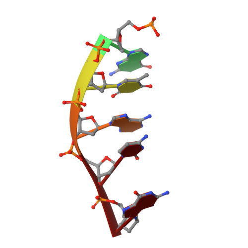
    Variolin B is a rare marine alkaloid that showed promising anti-cancer activity soon after its isolation. It acts as a cyclin-dependent kinase inhibitor, although the precise mechanism through which it exerts the cytotoxic effects is still unknown. The crystal structure of a variolin B bound to a DNA forming a pseudo-Holliday junction shows that this compound can also contribute, through intercalative binding, to either the formation or stabilization of multi-stranded DNA forms.


  • Organizational Affiliation

    Institute for Research in Biomedicine (IRB Barcelona), The Barcelona Institute of Science and Technology, Baldiri Reixac 10, 08028 Barcelona, Spain.


Macromolecules

Find similar nucleic acids by:  Sequence   |   3D Structure  

Entity ID: 1
MoleculeChains LengthOrganismImage
5'-D(*CP*GP*TP*AP*CP*G)-3'
A, B
6N/A
Sequence Annotations
Expand
  • Reference Sequence
Small Molecules
Ligands 4 Unique
IDChains Name / Formula / InChI Key2D Diagram3D Interactions
VAR
Query on VAR

Download Ideal Coordinates CCD File 
C [auth A],
D [auth A]
9-amino-5-(2-aminopyrimidin-4-yl)pyrido[3',2':4,5]pyrrolo[1,2-c]pyrimidin-4-ol
C14 H11 N7 O
RTHKPHCVZVYDFN-UHFFFAOYSA-N
NCO
Query on NCO

Download Ideal Coordinates CCD File 
E [auth A],
G [auth B],
H [auth B]
COBALT HEXAMMINE(III)
Co H18 N6
DYLMFCCYOUSRTK-UHFFFAOYSA-N
CO
Query on CO

Download Ideal Coordinates CCD File 
F [auth A]COBALT (II) ION
Co
XLJKHNWPARRRJB-UHFFFAOYSA-N
NA
Query on NA

Download Ideal Coordinates CCD File 
I [auth B]SODIUM ION
Na
FKNQFGJONOIPTF-UHFFFAOYSA-N
Experimental Data & Validation

Experimental Data

  • Method: X-RAY DIFFRACTION
  • Resolution: 1.40 Å
  • R-Value Free: 0.253 
  • R-Value Work: 0.208 
  • R-Value Observed: 0.210 
  • Space Group: C 2 2 2
Unit Cell:
Length ( Å )Angle ( ˚ )
a = 28.7α = 90
b = 53.4β = 90
c = 40.8γ = 90
Software Package:
Software NamePurpose
DNAdata collection
PHASERphasing
REFMACrefinement
XDSdata reduction
SCALAdata scaling

Structure Validation

View Full Validation Report


Ligand Structure Quality Assessment 


Entry History 

Deposition Data

Revision History  (Full details and data files)

  • Version 1.0: 2015-03-11
    Type: Initial release
  • Version 1.1: 2017-02-22
    Changes: Database references
  • Version 1.2: 2024-02-28
    Changes: Data collection, Database references, Derived calculations