4MGD

Crystal structure of hERa-LBD (Y537S) in complex with HPTE


Experimental Data Snapshot

  • Method: X-RAY DIFFRACTION
  • Resolution: 1.90 Å
  • R-Value Free: 0.234 
  • R-Value Work: 0.196 
  • R-Value Observed: 0.198 

wwPDB Validation   3D Report Full Report


Ligand Structure Quality Assessment 


This is version 1.3 of the entry. See complete history


Literature

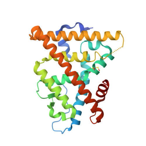
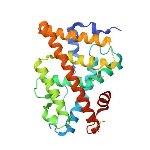
Structural and functional profiling of environmental ligands for estrogen receptors.

Delfosse, V.Grimaldi, M.Cavailles, V.Balaguer, P.Bourguet, W.

(2014) Environ Health Perspect 122: 1306-1313

  • DOI: https://doi.org/10.1289/ehp.1408453
  • Primary Citation of Related Structures:  
    4MG5, 4MG6, 4MG7, 4MG8, 4MG9, 4MGA, 4MGB, 4MGC, 4MGD

  • PubMed Abstract: 
  • Individuals are exposed daily to environmental pollutants that may act as endocrine-disrupting chemicals (EDCs), causing a range of developmental, reproductive, metabolic, or neoplastic diseases. With their mostly hydrophobic pocket that serves as a docking site for endogenous and exogenous ligands, nuclear receptors (NRs) can be primary targets of small molecule environmental contaminants ...

    Individuals are exposed daily to environmental pollutants that may act as endocrine-disrupting chemicals (EDCs), causing a range of developmental, reproductive, metabolic, or neoplastic diseases. With their mostly hydrophobic pocket that serves as a docking site for endogenous and exogenous ligands, nuclear receptors (NRs) can be primary targets of small molecule environmental contaminants. However, most of these compounds are chemically unrelated to natural hormones, so their binding modes and associated hormonal activities are hardly predictable.


    Organizational Affiliation

    Inserm (Institut national de la santé et de la recherche médicale) U1054, Montpellier, France.


Macromolecules
Find similar proteins by:  (by identity cutoff)  |  3D Structure
Entity ID: 1
MoleculeChainsSequence LengthOrganismDetailsImage
Estrogen receptor255Homo sapiensMutation(s): 1 
Gene Names: ESRESR1NR3A1
UniProt & NIH Common Fund Data Resources
Find proteins for P03372 (Homo sapiens)
Explore P03372 
Go to UniProtKB:  P03372
PHAROS:  P03372
Entity Groups  
Sequence Clusters30% Identity50% Identity70% Identity90% Identity95% Identity100% Identity
UniProt GroupP03372
Protein Feature View
Expand
  • Reference Sequence
Find similar proteins by:  (by identity cutoff)  |  3D Structure
Entity ID: 2
MoleculeChainsSequence LengthOrganismDetailsImage
Estrogen receptor255Homo sapiensMutation(s): 1 
Gene Names: ESRESR1NR3A1
UniProt & NIH Common Fund Data Resources
Find proteins for P03372 (Homo sapiens)
Explore P03372 
Go to UniProtKB:  P03372
PHAROS:  P03372
Entity Groups  
Sequence Clusters30% Identity50% Identity70% Identity90% Identity95% Identity100% Identity
UniProt GroupP03372
Protein Feature View
Expand
  • Reference Sequence

Find similar proteins by:  Sequence   |   3D Structure  

Entity ID: 3
MoleculeChainsSequence LengthOrganismDetailsImage
Nuclear receptor coactivator 1C [auth F],
D [auth G]
13Homo sapiensMutation(s): 0 
Gene Names: NCOA1BHLHE74SRC1
EC: 2.3.1.48
UniProt & NIH Common Fund Data Resources
Find proteins for Q15788 (Homo sapiens)
Explore Q15788 
Go to UniProtKB:  Q15788
PHAROS:  Q15788
Entity Groups  
Sequence Clusters30% Identity50% Identity70% Identity90% Identity95% Identity100% Identity
UniProt GroupQ15788
Protein Feature View
Expand
  • Reference Sequence
Small Molecules
Ligands 3 Unique
IDChainsName / Formula / InChI Key2D Diagram3D Interactions
27N
Query on 27N

Download Ideal Coordinates CCD File 
E [auth A],
F [auth B]
4,4'-(2,2,2-trichloroethane-1,1-diyl)diphenol
C14 H11 Cl3 O2
IUGDILGOLSSKNE-UHFFFAOYSA-N
 Ligand Interaction
GOL
Query on GOL

Download Ideal Coordinates CCD File 
G [auth B]GLYCEROL
C3 H8 O3
PEDCQBHIVMGVHV-UHFFFAOYSA-N
 Ligand Interaction
EDO
Query on EDO

Download Ideal Coordinates CCD File 
H [auth B]1,2-ETHANEDIOL
C2 H6 O2
LYCAIKOWRPUZTN-UHFFFAOYSA-N
 Ligand Interaction
Modified Residues  1 Unique
IDChainsTypeFormula2D DiagramParent
CSO
Query on CSO
A
L-PEPTIDE LINKINGC3 H7 N O3 SCYS
Experimental Data & Validation

Experimental Data

  • Method: X-RAY DIFFRACTION
  • Resolution: 1.90 Å
  • R-Value Free: 0.234 
  • R-Value Work: 0.196 
  • R-Value Observed: 0.198 
  • Space Group: P 1 21 1
Unit Cell:
Length ( Å )Angle ( ˚ )
a = 54.9α = 90
b = 81.88β = 110.54
c = 58.59γ = 90
Software Package:
Software NamePurpose
CBASSdata collection
PHENIXmodel building
PHENIXrefinement
XDSdata reduction
XSCALEdata scaling
PHENIXphasing

Structure Validation

View Full Validation Report


Ligand Structure Quality Assessment 


Entry History 

Deposition Data

Revision History  (Full details and data files)

  • Version 1.0: 2014-09-03
    Type: Initial release
  • Version 1.1: 2014-10-08
    Changes: Database references
  • Version 1.2: 2014-12-17
    Changes: Database references
  • Version 1.3: 2023-09-20
    Changes: Data collection, Database references, Derived calculations, Refinement description