4MDA

Structure of Mos1 transposase catalytic domain and Raltegravir with Mn


Experimental Data Snapshot

  • Method: X-RAY DIFFRACTION
  • Resolution: 1.70 Å
  • R-Value Free: 0.241 
  • R-Value Work: 0.197 
  • R-Value Observed: 0.199 

wwPDB Validation   3D Report Full Report


Ligand Structure Quality Assessment 


This is version 1.4 of the entry. See complete history


Literature

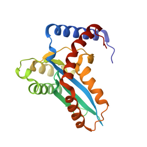
Structural Basis of Mos1 Transposase Inhibition by the Anti-retroviral Drug Raltegravir.

Wolkowicz, U.M.Morris, E.R.Robson, M.Trubitsyna, M.Richardson, J.M.

(2014) ACS Chem Biol 9: 743-751

  • DOI: https://doi.org/10.1021/cb400791u
  • Primary Citation of Related Structures:  
    4MDA, 4MDB

  • PubMed Abstract: 

    DNA transposases catalyze the movement of transposons around genomes by a cut-and-paste mechanism related to retroviral integration. Transposases and retroviral integrases share a common RNaseH-like domain with a catalytic DDE/D triad that coordinates the divalent cations required for DNA cleavage and integration. The anti-retroviral drugs Raltegravir and Elvitegravir inhibit integrases by displacing viral DNA ends from the catalytic metal ions. We demonstrate that Raltegravir, but not Elvitegravir, binds to Mos1 transposase in the presence of Mg(2+) or Mn(2+), without the requirement for transposon DNA, and inhibits transposon cleavage and DNA integration in biochemical assays. Crystal structures at 1.7 Å resolution show Raltegravir, in common with integrases, coordinating two Mg(2+) or Mn(2+) ions in the Mos1 active site. However, in the absence of transposon ends, the drug adopts an unusual, compact binding mode distinct from that observed in the active site of the prototype foamy virus integrase.


  • Organizational Affiliation

    School of Biological Sciences, University of Edinburgh , Mayfield Road, Edinburgh EH9 3JR, United Kingdom.


Macromolecules
Find similar proteins by:  (by identity cutoff)  |  3D Structure
Entity ID: 1
MoleculeChains Sequence LengthOrganismDetailsImage
Mariner Mos1 transposase225Drosophila mauritianaMutation(s): 1 
Gene Names: mariner\T
EC: 3.1
UniProt
Find proteins for Q7JQ07 (Drosophila mauritiana)
Explore Q7JQ07 
Go to UniProtKB:  Q7JQ07
Entity Groups  
Sequence Clusters30% Identity50% Identity70% Identity90% Identity95% Identity100% Identity
UniProt GroupQ7JQ07
Sequence Annotations
Expand
  • Reference Sequence
Small Molecules
Ligands 2 Unique
IDChains Name / Formula / InChI Key2D Diagram3D Interactions
RLT
Query on RLT

Download Ideal Coordinates CCD File 
D [auth A]N-(4-fluorobenzyl)-5-hydroxy-1-methyl-2-(1-methyl-1-{[(5-methyl-1,3,4-oxadiazol-2-yl)carbonyl]amino}ethyl)-6-oxo-1,6-di hydropyrimidine-4-carboxamide
C20 H21 F N6 O5
CZFFBEXEKNGXKS-UHFFFAOYSA-N
MN
Query on MN

Download Ideal Coordinates CCD File 
B [auth A],
C [auth A]
MANGANESE (II) ION
Mn
WAEMQWOKJMHJLA-UHFFFAOYSA-N
Experimental Data & Validation

Experimental Data

  • Method: X-RAY DIFFRACTION
  • Resolution: 1.70 Å
  • R-Value Free: 0.241 
  • R-Value Work: 0.197 
  • R-Value Observed: 0.199 
  • Space Group: P 41 21 2
Unit Cell:
Length ( Å )Angle ( ˚ )
a = 44.58α = 90
b = 44.58β = 90
c = 209.23γ = 90
Software Package:
Software NamePurpose
MOSFLMdata reduction
SCALAdata scaling
PHASERphasing
REFMACrefinement
PDB_EXTRACTdata extraction

Structure Validation

View Full Validation Report


Ligand Structure Quality Assessment 


Entry History 

Deposition Data

Revision History  (Full details and data files)

  • Version 1.0: 2014-01-22
    Type: Initial release
  • Version 1.1: 2014-02-05
    Changes: Database references
  • Version 1.2: 2014-04-02
    Changes: Database references
  • Version 1.3: 2014-05-07
    Changes: Other
  • Version 1.4: 2023-09-20
    Changes: Data collection, Database references, Derived calculations, Refinement description, Structure summary