4M9Z

Crystal structure of CED-4 bound CED-3 fragment


Experimental Data Snapshot

  • Method: X-RAY DIFFRACTION
  • Resolution: 3.40 Å
  • R-Value Free: 0.327 
  • R-Value Work: 0.270 
  • R-Value Observed: 0.272 

wwPDB Validation   3D Report Full Report


Ligand Structure Quality Assessment 


This is version 1.2 of the entry. See complete history


Literature

Mechanistic insights into CED-4-mediated activation of CED-3.

Huang, W.Jiang, T.Choi, W.Qi, S.Pang, Y.Hu, Q.Xu, Y.Gong, X.Jeffrey, P.D.Wang, J.Shi, Y.

(2013) Genes Dev 27: 2039-2048

  • DOI: https://doi.org/10.1101/gad.224428.113
  • Primary Citation of Related Structures:  
    4M9R, 4M9S, 4M9X, 4M9Y, 4M9Z

  • PubMed Abstract: 

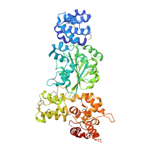
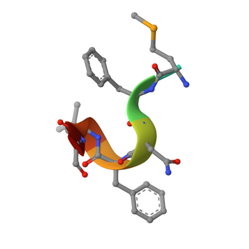
    Programmed cell death in Caenorhabditis elegans requires activation of the caspase CED-3, which strictly depends on CED-4. CED-4 forms an octameric apoptosome, which binds the CED-3 zymogen and facilitates its autocatalytic maturation. Despite recent advances, major questions remain unanswered. Importantly, how CED-4 recognizes CED-3 and how such binding facilitates CED-3 activation remain completely unknown. Here we demonstrate that the L2' loop of CED-3 directly binds CED-4 and plays a major role in the formation of an active CED-4-CED-3 holoenzyme. The crystal structure of the CED-4 apoptosome bound to the L2' loop fragment of CED-3, determined at 3.2 Å resolution, reveals specific interactions between a stretch of five hydrophobic amino acids from CED-3 and a shallow surface pocket within the hutch of the funnel-shaped CED-4 apoptosome. Structure-guided biochemical analysis confirms the functional importance of the observed CED-4-CED-3 interface. Structural analysis together with published evidence strongly suggest a working model in which two molecules of CED-3 zymogen, through specific recognition, are forced into the hutch of the CED-4 apoptosome, consequently undergoing dimerization and autocatalytic maturation. The mechanism of CED-3 activation represents a major revision of the prevailing model for initiator caspase activation.


  • Organizational Affiliation

    Ministry of Education Protein Science Laboratory, Center for Structural Biology, School of Life Sciences.


Macromolecules
Find similar proteins by:  (by identity cutoff)  |  3D Structure
Entity ID: 1
MoleculeChains Sequence LengthOrganismDetailsImage
Cell death protein 4A,
C [auth B],
E [auth C],
G [auth D]
549Caenorhabditis elegansMutation(s): 0 
Gene Names: ced-4C35D10.9
UniProt
Find proteins for P30429 (Caenorhabditis elegans)
Explore P30429 
Go to UniProtKB:  P30429
Entity Groups  
Sequence Clusters30% Identity50% Identity70% Identity90% Identity95% Identity100% Identity
UniProt GroupP30429
Sequence Annotations
Expand
  • Reference Sequence

Find similar proteins by:  Sequence   |   3D Structure  

Entity ID: 2
MoleculeChains Sequence LengthOrganismDetailsImage
CED-3 fragmentB [auth E],
D [auth F],
F [auth G],
H
8N/AMutation(s): 0 
Sequence Annotations
Expand
  • Reference Sequence
Experimental Data & Validation

Experimental Data

  • Method: X-RAY DIFFRACTION
  • Resolution: 3.40 Å
  • R-Value Free: 0.327 
  • R-Value Work: 0.270 
  • R-Value Observed: 0.272 
  • Space Group: P 4 21 2
Unit Cell:
Length ( Å )Angle ( ˚ )
a = 178.167α = 90
b = 178.167β = 90
c = 200.738γ = 90
Software Package:
Software NamePurpose
ADSCdata collection
PHASERphasing
PHENIXrefinement
HKL-2000data reduction
HKL-2000data scaling

Structure Validation

View Full Validation Report


Ligand Structure Quality Assessment 


Entry History 

Deposition Data

Revision History  (Full details and data files)

  • Version 1.0: 2013-10-23
    Type: Initial release
  • Version 1.1: 2023-11-08
    Changes: Data collection, Database references, Derived calculations, Refinement description
  • Version 1.2: 2023-12-06
    Changes: Data collection