4M38

Crystal structure of Trypanosoma brucei protein arginine methyltransferase 7 complex with AdoHcy and histone H4 peptide


Experimental Data Snapshot

  • Method: X-RAY DIFFRACTION
  • Resolution: 2.20 Å
  • R-Value Free: 0.230 
  • R-Value Work: 0.188 
  • R-Value Observed: 0.190 

wwPDB Validation   3D Report Full Report


Ligand Structure Quality Assessment 


This is version 1.1 of the entry. See complete history


Literature

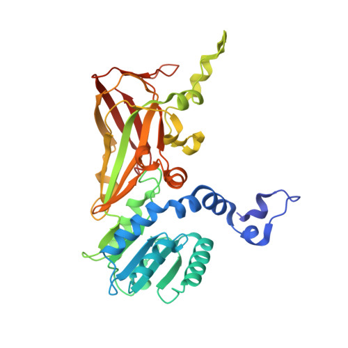
Structural determinants for the strict monomethylation activity by trypanosoma brucei protein arginine methyltransferase 7.

Wang, C.Zhu, Y.Caceres, T.B.Liu, L.Peng, J.Wang, J.Chen, J.Chen, X.Zhang, Z.Zuo, X.Gong, Q.Teng, M.Hevel, J.M.Wu, J.Shi, Y.

(2014) Structure 22: 756-768

  • DOI: https://doi.org/10.1016/j.str.2014.03.003
  • Primary Citation of Related Structures:  
    4M36, 4M37, 4M38

  • PubMed Abstract: 

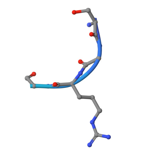
    Trypanosoma brucei protein arginine methyltransferase 7 (TbPRMT7) exclusively generates monomethylarginine (MMA), which directs biological consequences distinct from that of symmetric dimethylarginine (SDMA) and asymmetric dimethylarginine (ADMA). However, determinants controlling the strict monomethylation activity are unknown. We present the crystal structure of the TbPRMT7 active core in complex with S-adenosyl-L-homocysteine (AdoHcy) and a histone H4 peptide substrate. In the active site, residues E172, E181, and Q329 hydrogen bond the guanidino group of the target arginine and align the terminal guanidino nitrogen in a position suitable for nucleophilic attack on the methyl group of S-adenosyl-L-methionine (AdoMet). Structural comparisons and isothermal titration calorimetry data suggest that the TbPRMT7 active site is narrower than those of protein arginine dimethyltransferases, making it unsuitable to bind MMA in a manner that would support a second turnover, thus abolishing the production of SDMA and ADMA. Our results present the structural interpretations for the monomethylation activity of TbPRMT7.


  • Organizational Affiliation

    Hefei National Laboratory for Physical Sciences at Microscale and School of Life Sciences, University of Science and Technology of China, Anhui 230027, China.


Macromolecules
Find similar proteins by:  (by identity cutoff)  |  3D Structure
Entity ID: 1
MoleculeChains Sequence LengthOrganismDetailsImage
Protein arginine N-methyltransferase 7
A, B
343Trypanosoma brucei brucei TREU927Mutation(s): 0 
Gene Names: PRMT7Tb927.7.5490
EC: 2.1.1
UniProt
Find proteins for Q582G4 (Trypanosoma brucei brucei (strain 927/4 GUTat10.1))
Explore Q582G4 
Go to UniProtKB:  Q582G4
Entity Groups  
Sequence Clusters30% Identity50% Identity70% Identity90% Identity95% Identity100% Identity
UniProt GroupQ582G4
Sequence Annotations
Expand
  • Reference Sequence

Find similar proteins by:  Sequence   |   3D Structure  

Entity ID: 2
MoleculeChains Sequence LengthOrganismDetailsImage
Histone H4C [auth E],
D [auth F]
21Homo sapiensMutation(s): 0 
UniProt & NIH Common Fund Data Resources
Find proteins for P62805 (Homo sapiens)
Explore P62805 
Go to UniProtKB:  P62805
PHAROS:  P62805
Entity Groups  
Sequence Clusters30% Identity50% Identity70% Identity90% Identity95% Identity100% Identity
UniProt GroupP62805
Sequence Annotations
Expand
  • Reference Sequence
Small Molecules
Experimental Data & Validation

Experimental Data

  • Method: X-RAY DIFFRACTION
  • Resolution: 2.20 Å
  • R-Value Free: 0.230 
  • R-Value Work: 0.188 
  • R-Value Observed: 0.190 
  • Space Group: P 1 21 1
Unit Cell:
Length ( Å )Angle ( ˚ )
a = 66.696α = 90
b = 72.764β = 110.94
c = 72.565γ = 90
Software Package:
Software NamePurpose
HKL-2000data collection
MOLREPphasing
REFMACrefinement
HKL-2000data reduction
SCALAdata scaling

Structure Validation

View Full Validation Report


Ligand Structure Quality Assessment 


Entry History 

Deposition Data

Revision History  (Full details and data files)

  • Version 1.0: 2014-04-23
    Type: Initial release
  • Version 1.1: 2022-08-24
    Changes: Database references, Derived calculations