4LZ9 | pdb_00004lz9

Structure of MATE multidrug transporter DinF-BH in complex with R6G


Experimental Data Snapshot

  • Method: X-RAY DIFFRACTION
  • Resolution: 3.70 Å
  • R-Value Free: 
    0.300 (Depositor), 0.402 (DCC) 
  • R-Value Work: 
    0.290 (Depositor), 0.393 (DCC) 
  • R-Value Observed: 
    0.300 (Depositor) 

wwPDB Validation 3D Report Full Report

Validation slider image for 4LZ9

Ligand Structure Quality Assessment 


This is version 1.2 of the entry. See complete history

Literature

Structural insights into H(+)-coupled multidrug extrusion by a MATE transporter

Lu, M.Radchenko, M.Symersky, J.Nie, R.Guo, Y.

(2013) Nat Struct Mol Biol 20: 1310-1317

  • DOI: https://doi.org/10.1038/nsmb.2687
  • Primary Citation Related Structures: 
    4LZ6, 4LZ9

  • PubMed Abstract: 

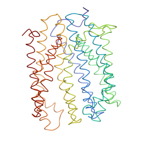
    Multidrug and toxic compound extrusion (MATE) transporters contribute to multidrug resistance by coupling the efflux of drugs to the influx of Na(+) or H(+). Known structures of Na(+)-coupled, extracellular-facing MATE transporters from the NorM subfamily revealed 12 membrane-spanning segments related by a quasi-two-fold rotational symmetry and a multidrug-binding cavity situated near the membrane surface. Here we report the crystal structure of an H(+)-coupled MATE transporter from Bacillus halodurans and the DinF subfamily at 3.2-Å resolution, unveiling a surprisingly asymmetric arrangement of 12 transmembrane helices. We also identified a membrane-embedded substrate-binding chamber by combining crystallographic and biochemical analyses. Our studies further suggested a direct competition between H(+) and substrate during DinF-mediated transport and implied how a MATE transporter alternates between its extracellular- and intracellular-facing conformations to propel multidrug extrusion. Collectively, our results demonstrated heretofore-unrecognized mechanistic diversity among MATE transporters.


  • Organizational Affiliation
    • Department of Biochemistry and Molecular Biology, Rosalind Franklin University of Medicine and Science, North Chicago, Illinois, USA.

Macromolecule Content 

  • Total Structure Weight: 49.06 kDa 
  • Atom Count: 479 
  • Modeled Residue Count: 446 
  • Deposited Residue Count: 446 
  • Unique protein chains: 1

Macromolecules

Find similar proteins by:|  3D Structure
Entity ID: 1
MoleculeChains  Sequence LengthOrganismDetailsImage
BH2163 protein446Halalkalibacterium halodurans C-125Mutation(s): 0 
Gene Names: BH2163
Membrane Entity: Yes 
UniProt
Find proteins for Q9KAX3 (Halalkalibacterium halodurans (strain ATCC BAA-125 / DSM 18197 / FERM 7344 / JCM 9153 / C-125))
Explore Q9KAX3 
Go to UniProtKB:  Q9KAX3
Entity Groups
Sequence Clusters30% Identity50% Identity70% Identity90% Identity95% Identity100% Identity
UniProt GroupQ9KAX3
Sequence Annotations
Expand
Reference Sequence

Small Molecules

Ligands 1 Unique
IDChains Name / Formula / InChI Key2D Diagram3D Interactions
RHQ

Query on RHQ


Download:Ideal Coordinates CCD File
B [auth A]RHODAMINE 6G
C28 H31 N2 O3
IWWWBRIIGAXLCJ-KRUMMXJUSA-O

Experimental Data & Validation

Experimental Data

  • Method: X-RAY DIFFRACTION
  • Resolution: 3.70 Å
  • R-Value Free:  0.300 (Depositor), 0.402 (DCC) 
  • R-Value Work:  0.290 (Depositor), 0.393 (DCC) 
  • R-Value Observed: 0.300 (Depositor) 
Space Group: P 21 21 21
Unit Cell:
Length ( Å )Angle ( ˚ )
a = 90.181α = 90
b = 93.887β = 90
c = 102.267γ = 90
Software Package:
Software NamePurpose
HKL-2000data collection
SHARPphasing
REFMACrefinement
HKL-2000data reduction
HKL-2000data scaling

Structure Validation

View Full Validation Report


Ligand Structure Quality Assessment 


Entry History 

Deposition Data

Revision History  (Full details and data files)

  • Version 1.0: 2013-10-23
    Type: Initial release
  • Version 1.1: 2013-11-20
    Changes: Database references
  • Version 1.2: 2024-03-20
    Changes: Data collection, Database references, Derived calculations