4LY8

dihydrodipicolinate synthase from C. jejuni with pyruvate bound to the active site


Experimental Data Snapshot

  • Method: X-RAY DIFFRACTION
  • Resolution: 1.70 Å
  • R-Value Free: 0.256 
  • R-Value Work: 0.209 
  • R-Value Observed: 0.211 

wwPDB Validation   3D Report Full Report


This is version 1.1 of the entry. See complete history


Literature

Tyrosine 110 Plays a Critical Role in Regulating the Allosteric Inhibition of Campylobacter jejuni Dihydrodipicolinate Synthase by Lysine.

Conly, C.J.Skovpen, Y.V.Li, S.Palmer, D.R.Sanders, D.A.

(2014) Biochemistry 53: 7396-7406

  • DOI: https://doi.org/10.1021/bi5012157
  • Primary Citation of Related Structures:  
    4LY8, 4M19, 4MLJ, 4MLR, 4R53

  • PubMed Abstract: 

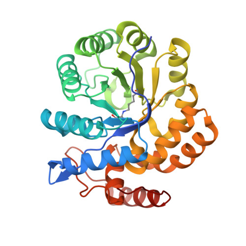
    Dihydrodipicolinate synthase (DHDPS), an enzyme found in most bacteria and plants, controls a critical step in the biosynthesis of l-lysine and meso-diaminopimelate, necessary components for bacterial cell wall biosynthesis. DHDPS catalyzes the condensation of pyruvate and (S)-aspartate-β-semialdehyde, forming an unstable product that is dehydrated to dihydrodipicolinate. The tetrameric enzyme is allosterically inhibited by l-lysine, and a better understanding of the allosteric inhibition mechanism is necessary for the design of potent antibacterial therapeutics. Here we describe the high-resolution crystal structures of DHDPS from Campylobacter jejuni with and without its inhibitor bound to the allosteric sites. These structures reveal a role for Y110 in the regulation of the allosteric inhibition by lysine. Mutation of Y110 to phenylalanine results in insensitivity to lysine inhibition, although the mutant crystal structure reveals that lysine does bind in the allosteric site. Comparison of the lysine-bound Y110F structure with wild-type structures reveals that key structural changes due to lysine binding are absent in this mutant.


  • Organizational Affiliation

    Department of Chemistry, University of Saskatchewan , 110 Science Place, Saskatoon, SK S7N 5C9, Canada.


Macromolecules
Find similar proteins by:  (by identity cutoff)  |  3D Structure
Entity ID: 1
MoleculeChains Sequence LengthOrganismDetailsImage
4-hydroxy-tetrahydrodipicolinate synthase
A, B, C, D
306Campylobacter jejuni subsp. jejuni NCTC 11168 = ATCC 700819Mutation(s): 0 
Gene Names: Cj0806dapA
EC: 4.3.3.7
UniProt
Find proteins for Q9PPB4 (Campylobacter jejuni subsp. jejuni serotype O:2 (strain ATCC 700819 / NCTC 11168))
Explore Q9PPB4 
Go to UniProtKB:  Q9PPB4
Entity Groups  
Sequence Clusters30% Identity50% Identity70% Identity90% Identity95% Identity100% Identity
UniProt GroupQ9PPB4
Sequence Annotations
Expand
  • Reference Sequence
Small Molecules
Ligands 5 Unique
IDChains Name / Formula / InChI Key2D Diagram3D Interactions
PG4
Query on PG4

Download Ideal Coordinates CCD File 
CA [auth C],
Q [auth A]
TETRAETHYLENE GLYCOL
C8 H18 O5
UWHCKJMYHZGTIT-UHFFFAOYSA-N
PGE
Query on PGE

Download Ideal Coordinates CCD File 
F [auth A]
FA [auth C]
H [auth A]
I [auth A]
JA [auth C]
F [auth A],
FA [auth C],
H [auth A],
I [auth A],
JA [auth C],
K [auth A],
LA [auth D],
T [auth B],
V [auth B],
X [auth B],
Y [auth B]
TRIETHYLENE GLYCOL
C6 H14 O4
ZIBGPFATKBEMQZ-UHFFFAOYSA-N
GOL
Query on GOL

Download Ideal Coordinates CCD File 
MA [auth D],
N [auth A],
R [auth A],
U [auth B],
Z [auth B]
GLYCEROL
C3 H8 O3
PEDCQBHIVMGVHV-UHFFFAOYSA-N
EDO
Query on EDO

Download Ideal Coordinates CCD File 
AA [auth B]
BA [auth B]
DA [auth C]
E [auth A]
EA [auth C]
AA [auth B],
BA [auth B],
DA [auth C],
E [auth A],
EA [auth C],
G [auth A],
GA [auth C],
HA [auth C],
IA [auth C],
J [auth A],
M [auth A],
NA [auth D],
O [auth A],
OA [auth D],
PA [auth D],
QA [auth D],
RA [auth D],
S [auth A],
SA [auth D],
TA [auth D],
W [auth B]
1,2-ETHANEDIOL
C2 H6 O2
LYCAIKOWRPUZTN-UHFFFAOYSA-N
ACT
Query on ACT

Download Ideal Coordinates CCD File 
KA [auth C],
L [auth A],
P [auth A]
ACETATE ION
C2 H3 O2
QTBSBXVTEAMEQO-UHFFFAOYSA-M
Modified Residues  1 Unique
IDChains TypeFormula2D DiagramParent
KPI
Query on KPI
A, B, C, D
L-PEPTIDE LINKINGC9 H16 N2 O4LYS
Experimental Data & Validation

Experimental Data

  • Method: X-RAY DIFFRACTION
  • Resolution: 1.70 Å
  • R-Value Free: 0.256 
  • R-Value Work: 0.209 
  • R-Value Observed: 0.211 
  • Space Group: P 21 21 21
Unit Cell:
Length ( Å )Angle ( ˚ )
a = 81.254α = 90
b = 101.404β = 90
c = 147.96γ = 90
Software Package:
Software NamePurpose
d*TREKdata scaling
d*TREKdata reduction
MOLREPphasing
PHENIXrefinement
PDB_EXTRACTdata extraction
MxDCdata collection

Structure Validation

View Full Validation Report


Entry History 

Deposition Data

Revision History  (Full details and data files)

  • Version 1.0: 2015-01-14
    Type: Initial release
  • Version 1.1: 2024-04-03
    Changes: Data collection, Database references, Derived calculations, Refinement description