


Murine IRGa6 bound to Toxoplasma ROP5B, a pseudokinase GDI
Reese, M.L., Shah, N., Boothroyd, J.C.To be published.
Experimental Data Snapshot
Starting Models: experimental
View more details
Entity ID: 1 | |||||
|---|---|---|---|---|---|
| Molecule | Chains | Sequence Length | Organism | Details | Image |
| Rhoptry protein 5B | 371 | Toxoplasma gondii | Mutation(s): 0 Gene Names: ROP5, ROP5B | 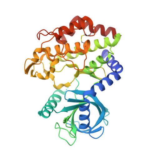 | |
UniProt | |||||
Entity Groups | |||||
| Sequence Clusters | 30% Identity50% Identity70% Identity90% Identity95% Identity100% Identity | ||||
| UniProt Group | F2YGR7 | ||||
Sequence AnnotationsExpand | |||||
Reference Sequence | |||||
Entity ID: 2 | |||||
|---|---|---|---|---|---|
| Molecule | Chains | Sequence Length | Organism | Details | Image |
| Interferon-inducible GTPase 1 | 423 | Mus musculus | Mutation(s): 0 Gene Names: Iigp1, Irga6 EC: 3.6.5 |  | |
UniProt | |||||
Entity Groups | |||||
| Sequence Clusters | 30% Identity50% Identity70% Identity90% Identity95% Identity100% Identity | ||||
| UniProt Group | Q9QZ85 | ||||
Sequence AnnotationsExpand | |||||
Reference Sequence | |||||
| Ligands 4 Unique | |||||
|---|---|---|---|---|---|
| ID | Chains | Name / Formula / InChI Key | 2D Diagram | 3D Interactions | |
| GDP Download:Ideal Coordinates CCD File | I [auth B] | GUANOSINE-5'-DIPHOSPHATE C10 H15 N5 O11 P2 QGWNDRXFNXRZMB-UUOKFMHZSA-N |  | ||
| ADP Download:Ideal Coordinates CCD File | C [auth A] | ADENOSINE-5'-DIPHOSPHATE C10 H15 N5 O10 P2 XTWYTFMLZFPYCI-KQYNXXCUSA-N |  | ||
| BME Download:Ideal Coordinates CCD File | D [auth A] | BETA-MERCAPTOETHANOL C2 H6 O S DGVVWUTYPXICAM-UHFFFAOYSA-N |  | ||
| EDO Download:Ideal Coordinates CCD File | E [auth A] F [auth A] G [auth A] H [auth A] J [auth B] | 1,2-ETHANEDIOL C2 H6 O2 LYCAIKOWRPUZTN-UHFFFAOYSA-N |  | ||
| Length ( Å ) | Angle ( ˚ ) |
|---|---|
| a = 52.6 | α = 99 |
| b = 54.7 | β = 106.6 |
| c = 85.6 | γ = 106.9 |
| Software Name | Purpose |
|---|---|
| PHASER | phasing |
| PHENIX | refinement |
| XSCALE | data scaling |
| PDB_EXTRACT | data extraction |


