4LV1 | pdb_00004lv1

AmpC beta-lactamase in complex with [1-(3-chlorophenyl)-1H-pyrazol-4-yl] boronic acid


Experimental Data Snapshot

  • Method: X-RAY DIFFRACTION
  • Resolution: 1.74 Å
  • R-Value Free: 
    0.195 (Depositor), 0.197 (DCC) 
  • R-Value Work: 
    0.170 (Depositor), 0.173 (DCC) 
  • R-Value Observed: 
    0.171 (Depositor) 

Starting Model: experimental
View more details

wwPDB Validation 3D Report Full Report

Validation slider image for 4LV1

Ligand Structure Quality Assessment 


This is version 1.4 of the entry. See complete history

Literature

Covalent docking of large libraries for the discovery of chemical probes.

London, N.Miller, R.M.Krishnan, S.Uchida, K.Irwin, J.J.Eidam, O.Gibold, L.Cimermancic, P.Bonnet, R.Shoichet, B.K.Taunton, J.

(2014) Nat Chem Biol 10: 1066-1072

  • DOI: https://doi.org/10.1038/nchembio.1666
  • Primary Citation Related Structures: 
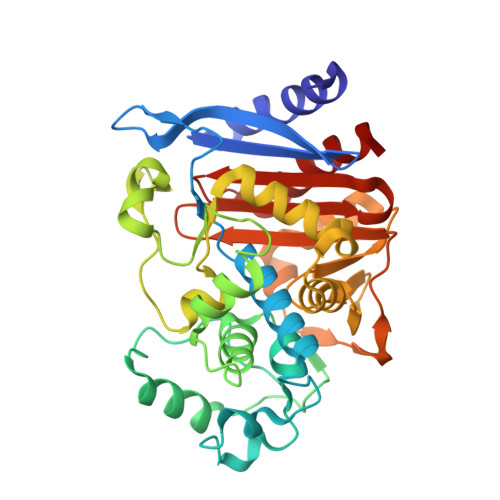
    4LV0, 4LV1, 4LV2, 4LV3, 4M8T

  • PubMed Abstract: 

    Chemical probes that form a covalent bond with a protein target often show enhanced selectivity, potency and utility for biological studies. Despite these advantages, protein-reactive compounds are usually avoided in high-throughput screening campaigns. Here we describe a general method (DOCKovalent) for screening large virtual libraries of electrophilic small molecules. We apply this method prospectively to discover reversible covalent fragments that target distinct protein nucleophiles, including the catalytic serine of AmpC β-lactamase and noncatalytic cysteines in RSK2, MSK1 and JAK3 kinases. We identify submicromolar to low-nanomolar hits with high ligand efficiency, cellular activity and selectivity, including what are to our knowledge the first reported reversible covalent inhibitors of JAK3. Crystal structures of inhibitor complexes with AmpC and RSK2 confirm the docking predictions and guide further optimization. As covalent virtual screening may have broad utility for the rapid discovery of chemical probes, we have made the method freely available through an automated web server (http://covalent.docking.org/).


  • Organizational Affiliation
    • Department of Pharmaceutical Chemistry, University of California-San Francisco, San Francisco, California, USA.

Macromolecule Content 

  • Total Structure Weight: 80.03 kDa 
  • Atom Count: 6,108 
  • Modeled Residue Count: 716 
  • Deposited Residue Count: 716 
  • Unique protein chains: 1

Macromolecules

Find similar proteins by:|  3D Structure
Entity ID: 1
MoleculeChains  Sequence LengthOrganismDetailsImage
Beta-lactamase
A, B
358Escherichia coli K-12Mutation(s): 0 
Gene Names: ampAampCb4150JW4111
EC: 3.5.2.6
UniProt
Find proteins for P00811 (Escherichia coli (strain K12))
Explore P00811 
Go to UniProtKB:  P00811
Entity Groups
Sequence Clusters30% Identity50% Identity70% Identity90% Identity95% Identity100% Identity
UniProt GroupP00811
Sequence Annotations
Expand
Reference Sequence

Experimental Data & Validation

Experimental Data

  • Method: X-RAY DIFFRACTION
  • Resolution: 1.74 Å
  • R-Value Free:  0.195 (Depositor), 0.197 (DCC) 
  • R-Value Work:  0.170 (Depositor), 0.173 (DCC) 
  • R-Value Observed: 0.171 (Depositor) 
Space Group: C 1 2 1
Unit Cell:
Length ( Å )Angle ( ˚ )
a = 118.58α = 90
b = 77.54β = 116.46
c = 97.64γ = 90
Software Package:
Software NamePurpose
XSCALEdata scaling
PHENIXrefinement
PDB_EXTRACTdata extraction

Structure Validation

View Full Validation Report


Ligand Structure Quality Assessment 


Entry History 

Deposition Data

Revision History  (Full details and data files)

  • Version 1.0: 2014-07-30
    Type: Initial release
  • Version 1.1: 2014-11-05
    Changes: Database references
  • Version 1.2: 2014-11-26
    Changes: Database references
  • Version 1.3: 2023-09-20
    Changes: Data collection, Database references, Derived calculations, Refinement description
  • Version 1.4: 2024-11-20
    Changes: Structure summary