Structural basis for DNA recognition and nuclease processing by the Mre11 homologue SbcD in double-strand breaks repair.
Liu, S., Tian, L.F., Liu, Y.P., An, X.M., Tang, Q., Yan, X.X., Liang, D.C.(2014) Acta Crystallogr D Biol Crystallogr 70: 299-309
- PubMed: 24531464 Search on PubMed
- DOI: https://doi.org/10.1107/S139900471302693X
- Primary Citation Related Structures:
4LTY, 4LU9, 4M0V - PubMed Abstract:
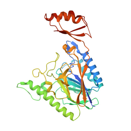
The Mre11 complex comprising meiotic recombination 11 (Mre11), Rad50 and Nijmegen breakage syndrome 1 (Nbs1) plays multiple important roles in the sensing, processing and repair of DNA double-strand breaks (DSBs). Here, crystal structures of the Escherichia coli Mre11 homologue SbcD and its Mn2+ complex are reported. Dimerization of SbcD depends on a four-helix bundle consisting of helices α2, α3, α2' and α3' of the two monomers, and the irregular and bent conformation of helices α3 and α3' in the SbcD dimer results in a dimeric arrangement that differs from those of previously reported Mre11 dimers. This finding indicates a distinct selectivity in DNA substrate recognition. The biochemical data combined with the crystal structures revealed that the SbcD monomer exhibits single-stranded DNA (ssDNA) endonuclease activity and double-stranded DNA (dsDNA) exonuclease activity on the addition of a high concentration of Mn2+. For the first time, atomic force microscopy analysis has been used to demonstrate that the SbcD monomer also possesses Mn2+-dependent dsDNA endonuclease activity. Loop β7-α6 of SbcD is likely to be a molecular switch and plays an important role in the regulation of substrate binding, catalytic reaction and state transitions. Based on structural and mutational analyses, a novel ssDNA-binding model of SbcD is proposed, providing insight into the catalytic mechanism of DSBs repair by the Mre11 complex.
- National Laboratory of Biomacromolecules, Institute of Biophysics, Chinese Academy of Sciences, Beijing 100101, People's Republic of China.
Organizational Affiliation: