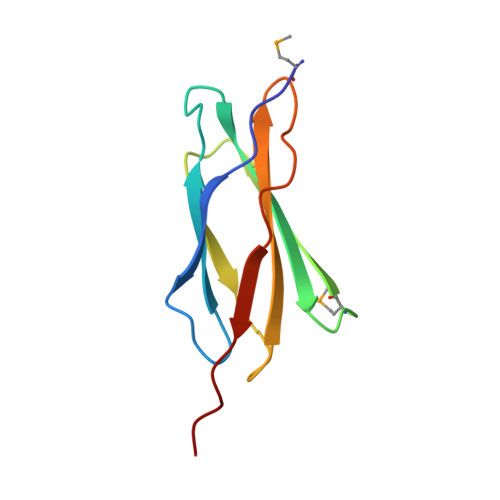
The structure of irisin reveals a novel intersubunit beta-sheet fibronectin type III (FNIII) dimer: implications for receptor activation.
Schumacher, M.A., Chinnam, N., Ohashi, T., Shah, R.S., Erickson, H.P.(2013) J Biol Chem 288: 33738-33744
- PubMed: 24114836 Search on PubMedSearch on PubMed Central
- DOI: https://doi.org/10.1074/jbc.M113.516641
- Primary Citation Related Structures:
4LSD - PubMed Abstract:
Irisin was recently identified as a putative myokine that is induced by exercise. Studies suggest that it is produced by cleavage of the FNDC5 (fibronectin domain-containing protein 5) receptor; irisin corresponds to the extracellular receptor ectodomain. Data suggesting that irisin stimulates white-to-brown fat conversion have led to the hypothesis that it does so by binding an unknown receptor, thus functioning as a myokine. As brown fat promotes energy dissipation, myokines that elicit the transformation of white to brown fat have potentially profound benefits in the treatment of obesity and metabolic disorders. Understanding the molecular basis for such exercise-induced phenomena is thus of considerable interest. Moreover, FNDC5-like receptors are highly conserved and have been shown to be critical for neuronal development. However, the structural and molecular mechanisms utilized by these proteins are currently unknown. Here, we describe the crystal structure and biochemical characterization of the FNDC5 ectodomain, corresponding to the irisin myokine. The 2.28 Å structure shows that irisin consists of an N-terminal fibronectin III (FNIII)-like domain attached to a flexible C-terminal tail. Strikingly, the FNIII-like domain forms a continuous intersubunit β-sheet dimer, previously unobserved for any FNIII protein. Biochemical data confirm that irisin is a dimer and that dimerization is unaffected by glycosylation. This finding suggests a possible mechanism for receptor activation by the irisin domain as a preformed myokine dimer ligand or as a paracrine or autocrine dimerization module on FNDC5-like receptors.
- Department of Biochemistry, Duke University School of Medicine, Durham, North Carolina 27710. Electronic address: maria.schumacher@duke.edu.
Organizational Affiliation: