4LQG | pdb_00004lqg

X-ray structure of human glutamate carboxypeptidase II (GCPII) in complex with a phosphoramidate inhibitor CTT1056


Experimental Data Snapshot

  • Method: X-RAY DIFFRACTION
  • Resolution: 1.77 Å
  • R-Value Free: 
    0.182 (Depositor) 
  • R-Value Work: 
    0.157 (Depositor) 
  • R-Value Observed: 
    0.159 (Depositor) 

wwPDB Validation 3D Report Full Report

Validation slider image for 4LQG

Ligand Structure Quality Assessment 


This is version 2.1 of the entry. See complete history

Literature

Structure-Activity Relationship of (18)F-Labeled Phosphoramidate Peptidomimetic Prostate-Specific Membrane Antigen (PSMA)-Targeted Inhibitor Analogues for PET Imaging of Prostate Cancer.

Dannoon, S.Ganguly, T.Cahaya, H.Geruntho, J.J.Galliher, M.S.Beyer, S.K.Choy, C.J.Hopkins, M.R.Regan, M.Blecha, J.E.Skultetyova, L.Drake, C.R.Jivan, S.Barinka, C.Jones, E.F.Berkman, C.E.VanBrocklin, H.F.

(2016) J Med Chem 59: 5684-5694

  • DOI: https://doi.org/10.1021/acs.jmedchem.5b01850
  • Primary Citation Related Structures: 
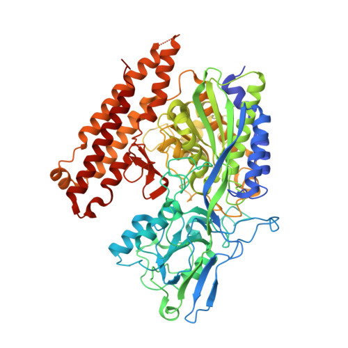
    4LQG

  • PubMed Abstract: 

    A series of phosphoramidate-based prostate specific membrane antigen (PSMA) inhibitors of increasing lipophilicity were synthesized (4, 5, and 6), and their fluorine-18 analogs were evaluated for use as positron emission tomography (PET) imaging agents for prostate cancer. To gain insight into their modes of binding, they were also cocrystallized with the extracellular domain of PSMA. All analogs exhibited irreversible binding to PSMA with IC50 values ranging from 0.4 to 1.3 nM. In vitro assays showed binding and rapid internalization (80-95%, 2 h) of the radiolabeled ligands in PSMA(+) cells. In vivo distribution demonstrated significant uptake in CWR22Rv1 (PSMA(+)) tumor, with tumor to blood ratios of 25.6:1, 63.6:1, and 69.6:1 for [(18)F]4, [(18)F]5, and [(18)F]6, respectively, at 2 h postinjection. Installation of aminohexanoic acid (AH) linkers in the phosphoramidate scaffold improved their PSMA binding and inhibition and was critical for achieving suitable in vivo imaging properties, positioning [(18)F]5 and [(18)F]6 as favorable candidates for future prostate cancer imaging clinical trials.


  • Organizational Affiliation
    • Department of Radiology and Biomedical Imaging, University of California-San Francisco , 185 Berry Street, San Francisco, California 94107, United States.

Macromolecule Content 

  • Total Structure Weight: 84.16 kDa 
  • Atom Count: 6,791 
  • Modeled Residue Count: 692 
  • Deposited Residue Count: 709 
  • Unique protein chains: 1

Macromolecules

Find similar proteins by:|  3D Structure
Entity ID: 1
MoleculeChains  Sequence LengthOrganismDetailsImage
Glutamate carboxypeptidase 2709Homo sapiensMutation(s): 0 
Gene Names: FOLHFOLH1GIG27NAALAD1PSMPSMA
EC: 3.4.17.21
UniProt & NIH Common Fund Data Resources
Find proteins for Q04609 (Homo sapiens)
Explore Q04609 
Go to UniProtKB:  Q04609
PHAROS:  Q04609
GTEx:  ENSG00000086205 
Entity Groups
Sequence Clusters30% Identity50% Identity70% Identity90% Identity95% Identity100% Identity
UniProt GroupQ04609
Glycosylation
Glycosylation Sites: 7Go to GlyGen: Q04609-1
Sequence Annotations
Expand
Reference Sequence

Oligosaccharides

Help  
Entity ID: 2
MoleculeChains Length2D Diagram GlycosylationD Interactions
2-acetamido-2-deoxy-beta-D-glucopyranose-(1-4)-2-acetamido-2-deoxy-beta-D-glucopyranose
B
2N-Glycosylation
Glycosylation Resources
GlyTouCan: G42666HT
GlyCosmos: G42666HT
GlyGen: G42666HT
Entity ID: 3
MoleculeChains Length2D Diagram GlycosylationD Interactions
2-acetamido-2-deoxy-beta-D-glucopyranose-(1-4)-[alpha-L-fucopyranose-(1-6)]2-acetamido-2-deoxy-beta-D-glucopyranose
C, D
3N-Glycosylation
Glycosylation Resources
GlyTouCan: G21290RB
GlyCosmos: G21290RB
GlyGen: G21290RB
Entity ID: 4
MoleculeChains Length2D Diagram GlycosylationD Interactions
beta-D-mannopyranose-(1-4)-2-acetamido-2-deoxy-beta-D-glucopyranose-(1-4)-2-acetamido-2-deoxy-beta-D-glucopyranose
E
3N-Glycosylation
Glycosylation Resources
GlyTouCan: G15407YE
GlyCosmos: G15407YE
GlyGen: G15407YE
Entity ID: 5
MoleculeChains Length2D Diagram GlycosylationD Interactions
alpha-D-mannopyranose-(1-3)-[alpha-D-mannopyranose-(1-6)]beta-D-mannopyranose-(1-4)-2-acetamido-2-deoxy-beta-D-glucopyranose-(1-4)-2-acetamido-2-deoxy-beta-D-glucopyranose
F
5N-Glycosylation
Glycosylation Resources
GlyTouCan: G22768VO
GlyCosmos: G22768VO
GlyGen: G22768VO

Small Molecules

Ligands 5 Unique
IDChains Name / Formula / InChI Key2D Diagram3D Interactions
CTW

Query on CTW


Download:Ideal Coordinates CCD File
M [auth A]N-(4-fluorobenzoyl)-L-gamma-glutamyl-5-{[(S)-{[(1S)-1,3-dicarboxypropyl]amino}(hydroxy)phosphoryl]oxy}-L-norvaline
C22 H29 F N3 O13 P
RHOKGKWGPOEIFI-JYJNAYRXSA-N
NAG

Query on NAG


Download:Ideal Coordinates CCD File
K [auth A],
L [auth A]
2-acetamido-2-deoxy-beta-D-glucopyranose
C8 H15 N O6
OVRNDRQMDRJTHS-FMDGEEDCSA-N
ZN

Query on ZN


Download:Ideal Coordinates CCD File
G [auth A],
H [auth A]
ZINC ION
Zn
PTFCDOFLOPIGGS-UHFFFAOYSA-N
CA

Query on CA


Download:Ideal Coordinates CCD File
I [auth A]CALCIUM ION
Ca
BHPQYMZQTOCNFJ-UHFFFAOYSA-N
CL

Query on CL


Download:Ideal Coordinates CCD File
J [auth A]CHLORIDE ION
Cl
VEXZGXHMUGYJMC-UHFFFAOYSA-M
Binding Affinity Annotations 
IDSourceBinding Affinity
CTW BindingDB:  4LQG IC50: 1300 (nM) from 1 assay(s)

Experimental Data & Validation

Experimental Data

  • Method: X-RAY DIFFRACTION
  • Resolution: 1.77 Å
  • R-Value Free:  0.182 (Depositor) 
  • R-Value Work:  0.157 (Depositor) 
  • R-Value Observed: 0.159 (Depositor) 
Space Group: I 2 2 2
Unit Cell:
Length ( Å )Angle ( ˚ )
a = 101.9α = 90
b = 130.233β = 90
c = 158.252γ = 90
Software Package:
Software NamePurpose
MAR345dtbdata collection
REFMACrefinement
XDSdata reduction
XDSdata scaling
REFMACphasing

Structure Validation

View Full Validation Report


Ligand Structure Quality Assessment 


Entry History 

Deposition Data

Revision History  (Full details and data files)

  • Version 1.0: 2014-12-31
    Type: Initial release
  • Version 1.1: 2016-06-15
    Changes: Database references
  • Version 1.2: 2016-06-29
    Changes: Database references
  • Version 1.3: 2016-07-06
    Changes: Database references
  • Version 1.4: 2017-11-15
    Changes: Refinement description
  • Version 2.0: 2020-07-29
    Type: Remediation
    Reason: Carbohydrate remediation
    Changes: Atomic model, Data collection, Database references, Derived calculations, Structure summary
  • Version 2.1: 2024-11-27
    Changes: Data collection, Database references, Structure summary