4LQE

Crystal Structure of MepB


Experimental Data Snapshot

  • Method: X-RAY DIFFRACTION
  • Resolution: 2.10 Å
  • R-Value Free: 0.279 
  • R-Value Work: 0.236 
  • R-Value Observed: 0.238 

wwPDB Validation   3D Report Full Report


This is version 1.1 of the entry. See complete history


Literature

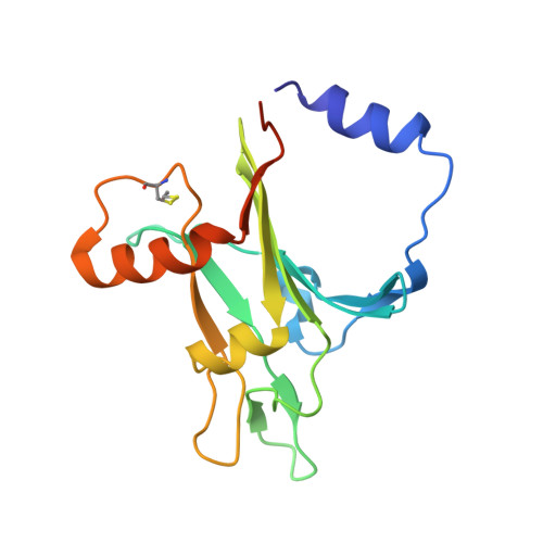
Structural characterization of MepB from Staphylococcus aureus reveals homology to endonucleases.

Agah, S.Poulos, S.Banchs, C.Faham, S.

(2014) Protein Sci 23: 594-602

  • DOI: https://doi.org/10.1002/pro.2438
  • Primary Citation of Related Structures:  
    4LQE

  • PubMed Abstract: 

    The MepRAB operon in Staphylococcus aureus has been identified to play a role in drug resistance. Although the functions of MepA and MepR are known, little information is available on the function of MepB. Here we report the X-ray structure of MepB to 2.1 Å revealing its structural similarity to the PD-(D/E)XK family of endonucleases. We further show that MepB binds DNA and RNA, with a higher affinity towards RNA and single stranded DNA than towards double stranded DNA. Notably, the PD-(D/E)XK catalytic active site residues are not conserved in MepB. MepB's association with a drug resistance operon suggests that it plays a role in responding to antimicrobials. This role is likely carried out through MepB's interactions with nucleic acids.


  • Organizational Affiliation

    Department of Molecular Physiology and Biological Physics, University of Virginia School of Medicine, Charlottesville, Virginia.


Macromolecules
Find similar proteins by:  (by identity cutoff)  |  3D Structure
Entity ID: 1
MoleculeChains Sequence LengthOrganismDetailsImage
MepB160Staphylococcus aureus subsp. aureus Mu50Mutation(s): 0 
Gene Names: MepBSAV0335
UniProt
Find proteins for A0A0H3JPI4 (Staphylococcus aureus (strain Mu50 / ATCC 700699))
Explore A0A0H3JPI4 
Go to UniProtKB:  A0A0H3JPI4
Entity Groups  
Sequence Clusters30% Identity50% Identity70% Identity90% Identity95% Identity100% Identity
UniProt GroupA0A0H3JPI4
Sequence Annotations
Expand
  • Reference Sequence
Small Molecules
Ligands 1 Unique
IDChains Name / Formula / InChI Key2D Diagram3D Interactions
SO4
Query on SO4

Download Ideal Coordinates CCD File 
B [auth A]SULFATE ION
O4 S
QAOWNCQODCNURD-UHFFFAOYSA-L
Modified Residues  1 Unique
IDChains TypeFormula2D DiagramParent
CME
Query on CME
A
L-PEPTIDE LINKINGC5 H11 N O3 S2CYS
Experimental Data & Validation

Experimental Data

  • Method: X-RAY DIFFRACTION
  • Resolution: 2.10 Å
  • R-Value Free: 0.279 
  • R-Value Work: 0.236 
  • R-Value Observed: 0.238 
  • Space Group: I 2 2 2
Unit Cell:
Length ( Å )Angle ( ˚ )
a = 44.645α = 90
b = 91.139β = 90
c = 100.004γ = 90
Software Package:
Software NamePurpose
CRANKphasing
REFMACrefinement
HKL-2000data reduction
SCALEPACKdata scaling

Structure Validation

View Full Validation Report


Entry History 

Deposition Data

Revision History  (Full details and data files)

  • Version 1.0: 2014-03-12
    Type: Initial release
  • Version 1.1: 2014-05-07
    Changes: Database references