Structures of oncogenic, suppressor and rescued p53 core-domain variants: mechanisms of mutant p53 rescue.
Wallentine, B.D., Wang, Y., Tretyachenko-Ladokhina, V., Tan, M., Senear, D.F., Luecke, H.(2013) Acta Crystallogr D Biol Crystallogr 69: 2146-2156
- PubMed: 24100332 Search on PubMedSearch on PubMed Central
- DOI: https://doi.org/10.1107/S0907444913020830
- Primary Citation Related Structures:
4KVP, 4LO9, 4LOE, 4LOF - PubMed Abstract:
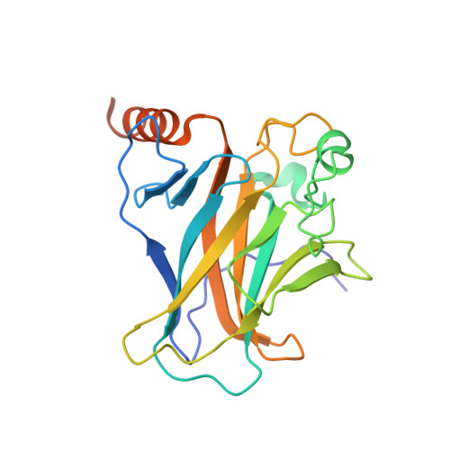
To gain insights into the mechanisms by which certain second-site suppressor mutations rescue the function of a significant number of cancer mutations of the tumor suppressor protein p53, X-ray crystallographic structures of four p53 core-domain variants were determined. These include an oncogenic mutant, V157F, two single-site suppressor mutants, N235K and N239Y, and the rescued cancer mutant V157F/N235K/N239Y. The V157F mutation substitutes a smaller hydrophobic valine with a larger hydrophobic phenylalanine within strand S4 of the hydrophobic core. The structure of this cancer mutant shows no gross structural changes in the overall fold of the p53 core domain, only minor rearrangements of side chains within the hydrophobic core of the protein. Based on biochemical analysis, these small local perturbations induce instability in the protein, increasing the free energy by 3.6 kcal mol(-1) (15.1 kJ mol(-1)). Further biochemical evidence shows that each suppressor mutation, N235K or N239Y, acts individually to restore thermodynamic stability to V157F and that both together are more effective than either alone. All rescued mutants were found to have wild-type DNA-binding activity when assessed at a permissive temperature, thus pointing to thermodynamic stability as the critical underlying variable. Interestingly, thermodynamic analysis shows that while N239Y demonstrates stabilization of the wild-type p53 core domain, N235K does not. These observations suggest distinct structural mechanisms of rescue. A new salt bridge between Lys235 and Glu198, found in both the N235K and rescued cancer mutant structures, suggests a rescue mechanism that relies on stabilizing the β-sandwich scaffold. On the other hand, the substitution N239Y creates an advantageous hydrophobic contact between the aromatic ring of this tyrosine and the adjacent Leu137. Surprisingly, the rescued cancer mutant shows much larger structural deviations than the cancer mutant alone when compared with wild-type p53. These suppressor mutations appear to rescue p53 function by creating novel intradomain interactions that stabilize the core domain, allowing compensation for the destabilizing V157F mutation.
- Department of Molecular Biology and Biochemistry, University of California, Irvine, Irvine, CA 92697, USA.
Organizational Affiliation: