4LNQ | pdb_00004lnq

Crystal structure of Ifi202 HINa domain in complex with 20bp dsDNA


Experimental Data Snapshot

  • Method: X-RAY DIFFRACTION
  • Resolution: 2.00 Å
  • R-Value Free: 
    0.234 (Depositor), 0.228 (DCC) 
  • R-Value Work: 
    0.200 (Depositor), 0.195 (DCC) 
  • R-Value Observed: 
    0.202 (Depositor) 

Starting Model: experimental
View more details

wwPDB Validation 3D Report Full Report

Validation slider image for 4LNQ

This is version 1.2 of the entry. See complete history

Literature

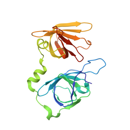
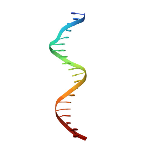
Structural mechanism of DNA recognition by the p202 HINa domain: insights into the inhibition of Aim2-mediated inflammatory signalling.

Li, H.Wang, J.Wang, J.Cao, L.S.Wang, Z.X.Wu, J.W.

(2014) Acta Crystallogr Sect F Struct Biol Cryst Commun 70: 21-29

  • DOI: https://doi.org/10.1107/S2053230X1303135X
  • Primary Citation Related Structures: 
    4LNQ

  • PubMed Abstract: 

    The HIN-200 family of proteins play significant roles in inflammation-related processes. Among them, AIM2 (absent in melanoma 2) and IFI16 (γ-interferon-inducible protein 16) recognize double-stranded DNA to initiate inflammatory responses. In contrast, p202, a mouse interferon-inducible protein containing two HIN domains (HINa and HINb), has been reported to inhibit Aim2-mediated inflammatory signalling in mouse. To understand the inhibitory mechanism, the crystal structure of the p202 HINa domain in complex with a 20 bp DNA was determined, in which p202 HINa nonspecifically recognizes both strands of DNA through electrostatic attraction. The p202 HINa domain binds DNA more tightly than does AIM2 HIN, and the DNA-binding mode of p202 HINa is different from that of the AIM2 HIN and IFI16 HINb domains. These results, together with the reported data on p202 HINb, lead to an interaction model for full-length p202 and dsDNA which provides a conceivable mechanism for the negative regulation of Aim2 inflammasome activation by p202.


  • Organizational Affiliation
    • MOE Key Laboratory of Protein Science and Tsinghua-Peking Center for Life Sciences, School of Life Sciences, Tsinghua University, Beijing 100084, People's Republic of China.

Macromolecule Content 

  • Total Structure Weight: 57.46 kDa 
  • Atom Count: 4,264 
  • Modeled Residue Count: 427 
  • Deposited Residue Count: 434 
  • Unique protein chains: 1
  • Unique nucleic acid chains: 1

Macromolecules


Find similar proteins by:|  3D Structure
Entity ID: 1
MoleculeChains  Sequence LengthOrganismDetailsImage
Interferon-activable protein 202
A, B
197Mus musculusMutation(s): 4 
Gene Names: Ifi202Ifi202aIfi202b
UniProt
Find proteins for Q9R002 (Mus musculus)
Explore Q9R002 
Go to UniProtKB:  Q9R002
Entity Groups
Sequence Clusters30% Identity50% Identity70% Identity90% Identity95% Identity100% Identity
UniProt GroupQ9R002
Sequence Annotations
Expand
Reference Sequence
Find similar nucleic acids by:  Sequence
Entity ID: 2
MoleculeChains LengthOrganismImage
20bp DNA
C, D
20N/A
Sequence Annotations
Expand
Reference Sequence

Experimental Data & Validation

Experimental Data

  • Method: X-RAY DIFFRACTION
  • Resolution: 2.00 Å
  • R-Value Free:  0.234 (Depositor), 0.228 (DCC) 
  • R-Value Work:  0.200 (Depositor), 0.195 (DCC) 
  • R-Value Observed: 0.202 (Depositor) 
Space Group: P 21 21 2
Unit Cell:
Length ( Å )Angle ( ˚ )
a = 95.353α = 90
b = 105.566β = 90
c = 65.143γ = 90
Software Package:
Software NamePurpose
HKL-2000data collection
PHASERphasing
PHENIXrefinement
HKL-2000data reduction
HKL-2000data scaling

Structure Validation

View Full Validation Report


Entry History 

Deposition Data

Revision History  (Full details and data files)

  • Version 1.0: 2014-01-08
    Type: Initial release
  • Version 1.1: 2014-02-12
    Changes: Database references
  • Version 1.2: 2023-09-20
    Changes: Data collection, Database references, Refinement description