4LHU | pdb_00004lhu

Crystal Structure of 9C2 TCR bound to CD1d


Experimental Data Snapshot

  • Method: X-RAY DIFFRACTION
  • Resolution: 2.87 Å
  • R-Value Free: 
    0.243 (Depositor), 0.262 (DCC) 
  • R-Value Work: 
    0.206 (Depositor), 0.223 (DCC) 
  • R-Value Observed: 
    0.208 (Depositor) 

Starting Models: experimental
View more details

wwPDB Validation 3D Report Full Report

Validation slider image for 4LHU

Ligand Structure Quality Assessment 


This is version 2.2 of the entry. See complete history

Literature

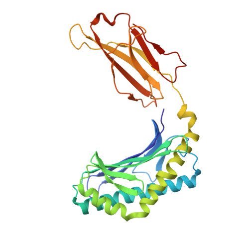
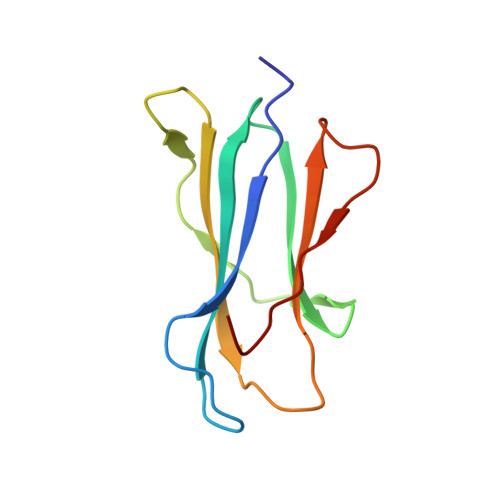
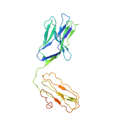
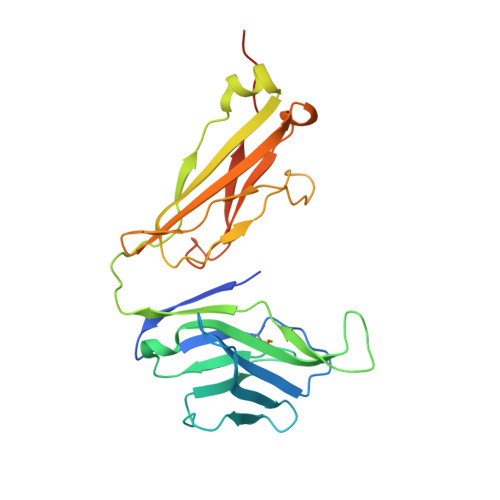
CD1d-lipid antigen recognition by the gamma delta TCR.

Uldrich, A.P.Le Nours, J.Pellicci, D.G.Gherardin, N.A.McPherson, K.G.Lim, R.T.Patel, O.Beddoe, T.Gras, S.Rossjohn, J.Godfrey, D.I.

(2013) Nat Immunol 14: 1137-1145

  • DOI: https://doi.org/10.1038/ni.2713
  • Primary Citation Related Structures: 
    4LFH, 4LHU

  • PubMed Abstract: 

    The T cell repertoire comprises αβ and γδ T cell lineages. Although it is established how αβ T cell antigen receptors (TCRs) interact with antigen presented by antigen-presenting molecules, this is unknown for γδ TCRs. We describe a population of human Vδ1(+) γδ T cells that exhibit autoreactivity to CD1d and provide a molecular basis for how a γδ TCR binds CD1d-α-galactosylceramide (α-GalCer). The γδ TCR docked orthogonally, over the A' pocket of CD1d, in which the Vδ1-chain, and in particular the germ line-encoded CDR1δ loop, dominated interactions with CD1d. The TCR γ-chain sat peripherally to the interface, with the CDR3γ loop representing the principal determinant for α-GalCer specificity. Accordingly, we provide insight into how a γδ TCR binds specifically to a lipid-loaded antigen-presenting molecule.


  • Organizational Affiliation
    • 1] Department of Microbiology and Immunology, Peter Doherty Institute for Infection and Immunity, University of Melbourne, Parkville, Victoria, Australia. [2].

Macromolecule Content 

  • Total Structure Weight: 101.31 kDa 
  • Atom Count: 6,749 
  • Modeled Residue Count: 801 
  • Deposited Residue Count: 865 
  • Unique protein chains: 4

Macromolecules

Find similar proteins by:|  3D Structure
Entity ID: 1
MoleculeChains  Sequence LengthOrganismDetailsImage
Antigen-presenting glycoprotein CD1d278Homo sapiensMutation(s): 0 
Gene Names: CD1D
UniProt & NIH Common Fund Data Resources
Find proteins for P15813 (Homo sapiens)
Explore P15813 
Go to UniProtKB:  P15813
PHAROS:  P15813
GTEx:  ENSG00000158473 
Entity Groups
Sequence Clusters30% Identity50% Identity70% Identity90% Identity95% Identity100% Identity
UniProt GroupP15813
Glycosylation
Glycosylation Sites: 3Go to GlyGen: P15813-1
Sequence Annotations
Expand
Reference Sequence
Find similar proteins by:|  3D Structure
Entity ID: 2
MoleculeChains  Sequence LengthOrganismDetailsImage
Beta-2-microglobulin100Homo sapiensMutation(s): 0 
Gene Names: B2MCDABP0092HDCMA22P
UniProt & NIH Common Fund Data Resources
Find proteins for P61769 (Homo sapiens)
Explore P61769 
Go to UniProtKB:  P61769
PHAROS:  P61769
GTEx:  ENSG00000166710 
Entity Groups
Sequence Clusters30% Identity50% Identity70% Identity90% Identity95% Identity100% Identity
UniProt GroupP61769
Sequence Annotations
Expand
Reference Sequence
Find similar proteins by:|  3D Structure
Entity ID: 3
MoleculeChains  Sequence LengthOrganismDetailsImage
9C2 TCR delta chainC [auth D]236Homo sapiensMutation(s): 0 
UniProt
Find proteins for Q6PJ56 (Homo sapiens)
Explore Q6PJ56 
Go to UniProtKB:  Q6PJ56
Entity Groups
Sequence Clusters30% Identity50% Identity70% Identity90% Identity95% Identity100% Identity
UniProt GroupQ6PJ56
Glycosylation
Glycosylation Sites: 1
Sequence Annotations
Expand
Reference Sequence
Find similar proteins by:|  3D Structure
Entity ID: 4
MoleculeChains  Sequence LengthOrganismDetailsImage
9C2 TCR gamma chainD [auth G]251Homo sapiensMutation(s): 0 
UniProt
Find proteins for P0CF51 (Homo sapiens)
Explore P0CF51 
Go to UniProtKB:  P0CF51
Entity Groups
Sequence Clusters30% Identity50% Identity70% Identity90% Identity95% Identity100% Identity
UniProt GroupP0CF51
Sequence Annotations
Expand
Reference Sequence

Oligosaccharides

Help  
Entity ID: 5
MoleculeChains Length2D Diagram GlycosylationD Interactions
2-acetamido-2-deoxy-beta-D-glucopyranose-(1-4)-2-acetamido-2-deoxy-beta-D-glucopyranoseE [auth C],
F [auth E]
2N-Glycosylation
Glycosylation Resources
GlyTouCan: G42666HT
GlyCosmos: G42666HT
GlyGen: G42666HT
Entity ID: 6
MoleculeChains Length2D Diagram GlycosylationD Interactions
beta-D-mannopyranose-(1-4)-2-acetamido-2-deoxy-beta-D-glucopyranose-(1-4)-2-acetamido-2-deoxy-beta-D-glucopyranoseG [auth F]3N-Glycosylation
Glycosylation Resources
GlyTouCan: G15407YE
GlyCosmos: G15407YE
GlyGen: G15407YE

Small Molecules

Ligands 5 Unique
IDChains Name / Formula / InChI Key2D Diagram3D Interactions
JLS

Query on JLS


Download:Ideal Coordinates CCD File
K [auth A](15Z)-N-[(2S,3S,4R)-1-(alpha-D-galactopyranosyloxy)-3,4-dihydroxyoctadecan-2-yl]tetracos-15-enamide
C48 H93 N O9
QMLKBWVXCMTWOZ-WWHSRKRPSA-N
NAG

Query on NAG


Download:Ideal Coordinates CCD File
H [auth A]2-acetamido-2-deoxy-beta-D-glucopyranose
C8 H15 N O6
OVRNDRQMDRJTHS-FMDGEEDCSA-N
BMA

Query on BMA


Download:Ideal Coordinates CCD File
I [auth A]beta-D-mannopyranose
C6 H12 O6
WQZGKKKJIJFFOK-RWOPYEJCSA-N
CL

Query on CL


Download:Ideal Coordinates CCD File
L [auth G]CHLORIDE ION
Cl
VEXZGXHMUGYJMC-UHFFFAOYSA-M
MG

Query on MG


Download:Ideal Coordinates CCD File
J [auth A]MAGNESIUM ION
Mg
JLVVSXFLKOJNIY-UHFFFAOYSA-N

Experimental Data & Validation

Experimental Data

  • Method: X-RAY DIFFRACTION
  • Resolution: 2.87 Å
  • R-Value Free:  0.243 (Depositor), 0.262 (DCC) 
  • R-Value Work:  0.206 (Depositor), 0.223 (DCC) 
  • R-Value Observed: 0.208 (Depositor) 
Space Group: C 2 2 21
Unit Cell:
Length ( Å )Angle ( ˚ )
a = 131.62α = 90
b = 152.69β = 90
c = 135.2γ = 90
Software Package:
Software NamePurpose
SCALAdata scaling
PHASERphasing
BUSTER-TNTrefinement
PDB_EXTRACTdata extraction
Blu-Icedata collection
XDSdata reduction
BUSTERrefinement

Structure Validation

View Full Validation Report


Ligand Structure Quality Assessment 


Entry History 

Deposition Data

Revision History  (Full details and data files)

  • Version 1.0: 2013-10-02
    Type: Initial release
  • Version 1.1: 2013-10-16
    Changes: Database references
  • Version 1.2: 2013-11-06
    Changes: Database references
  • Version 2.0: 2020-07-29
    Type: Remediation
    Reason: Carbohydrate remediation
    Changes: Advisory, Atomic model, Data collection, Database references, Derived calculations, Structure summary
  • Version 2.1: 2023-09-20
    Changes: Advisory, Data collection, Database references, Refinement description, Structure summary
  • Version 2.2: 2024-11-27
    Changes: Structure summary