


The Crystal Structure of Human Methyltransferase-Like Protein 21D in Complex with SAM
Zeng, H., Dong, A., Fenner, M., Bountra, C., Arrowsmith, C.H., Edwards, A.M., Brown, P.J., Wu, H., Structural Genomics Consortium (SGC)To be published.
Experimental Data Snapshot
Starting Model: experimental
View more details
Entity ID: 1 | |||||
|---|---|---|---|---|---|
| Molecule | Chains | Sequence Length | Organism | Details | Image |
| Protein-lysine methyltransferase METTL21D | 224 | Homo sapiens | Mutation(s): 0 Gene Names: METTL21D, C14orf138 EC: 2.1.1 | 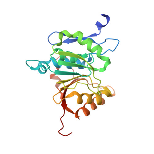 | |
UniProt & NIH Common Fund Data Resources | |||||
PHAROS: Q9H867 GTEx: ENSG00000100483 | |||||
Entity Groups | |||||
| Sequence Clusters | 30% Identity50% Identity70% Identity90% Identity95% Identity100% Identity | ||||
| UniProt Group | Q9H867 | ||||
Sequence AnnotationsExpand | |||||
Reference Sequence | |||||
| Ligands 3 Unique | |||||
|---|---|---|---|---|---|
| ID | Chains | Name / Formula / InChI Key | 2D Diagram | 3D Interactions | |
| SAM Download:Ideal Coordinates CCD File | D [auth A], O [auth B], Z [auth C] | S-ADENOSYLMETHIONINE C15 H22 N6 O5 S MEFKEPWMEQBLKI-FCKMPRQPSA-N |  | ||
| EDO Download:Ideal Coordinates CCD File | E [auth A], P [auth B] | 1,2-ETHANEDIOL C2 H6 O2 LYCAIKOWRPUZTN-UHFFFAOYSA-N |  | ||
| UNX Download:Ideal Coordinates CCD File | AA [auth C] BA [auth C] CA [auth C] DA [auth C] EA [auth C] | UNKNOWN ATOM OR ION X |  | ||
| Length ( Å ) | Angle ( ˚ ) |
|---|---|
| a = 47.783 | α = 90 |
| b = 100.052 | β = 101.25 |
| c = 96.193 | γ = 90 |
| Software Name | Purpose |
|---|---|
| SCALEPACK | data scaling |
| REFMAC | refinement |
| PDB_EXTRACT | data extraction |
| SBC-Collect | data collection |
| HKL-3000 | data reduction |
| HKL-3000 | data scaling |
| PHASER | phasing |


