


Structures of a GH131 beta-Glucanase Catalytic Domain from Podospora anserina in Complex with Cellotriose
Jiang, T., Chan, H.C., Huang, C.H., Ko, T.P., Huang, T.Y., Liu, J.R., Guo, R.T.To be published.
Experimental Data Snapshot
wwPDB Validation 3D Report Full Report

Entity ID: 1 | |||||
|---|---|---|---|---|---|
| Molecule | Chains | Sequence Length | Organism | Details | Image |
| Beta-glucanase | 252 | Podospora anserina | Mutation(s): 0 | 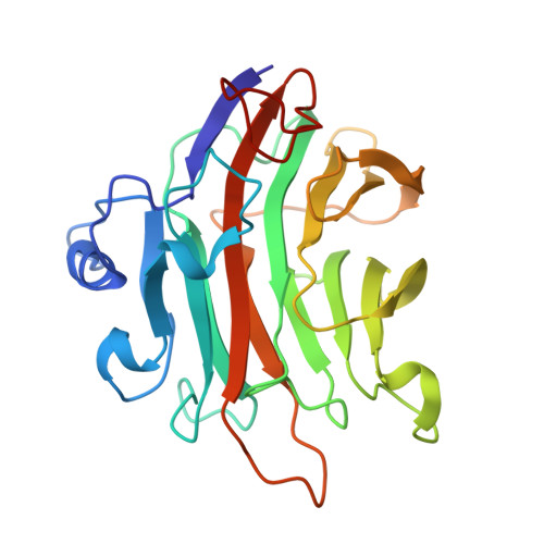 | |
UniProt | |||||
Entity Groups | |||||
| Sequence Clusters | 30% Identity50% Identity70% Identity90% Identity95% Identity100% Identity | ||||
| UniProt Group | J7K096 | ||||
Sequence AnnotationsExpand | |||||
Reference Sequence | |||||
| Length ( Å ) | Angle ( ˚ ) |
|---|---|
| a = 54.821 | α = 81.61 |
| b = 62.167 | β = 75.01 |
| c = 79.154 | γ = 77.15 |
| Software Name | Purpose |
|---|---|
| HKL-2000 | data collection |
| OASIS | model building |
| CNS | refinement |
| HKL-2000 | data reduction |
| HKL-2000 | data scaling |
| OASIS | phasing |


