4LCD | pdb_00004lcd

Structure of an Rsp5xUbxSna3 complex: Mechanism of ubiquitin ligation and lysine prioritization by a HECT E3


Experimental Data Snapshot

  • Method: X-RAY DIFFRACTION
  • Resolution: 3.10 Å
  • R-Value Free: 
    0.299 (Depositor), 0.297 (DCC) 
  • R-Value Work: 
    0.251 (Depositor), 0.252 (DCC) 
  • R-Value Observed: 
    0.253 (Depositor) 

wwPDB Validation 3D Report Full Report

Validation slider image for 4LCD

This is version 1.2 of the entry. See complete history

Literature

Mechanism of ubiquitin ligation and lysine prioritization by a HECT E3.

Kamadurai, H.B.Qiu, Y.Deng, A.Harrison, J.S.Macdonald, C.Actis, M.Rodrigues, P.Miller, D.J.Souphron, J.Lewis, S.M.Kurinov, I.Fujii, N.Hammel, M.Piper, R.Kuhlman, B.Schulman, B.A.

(2013) Elife 2: e00828-e00828

  • DOI: https://doi.org/10.7554/eLife.00828
  • Primary Citation Related Structures: 
    4LCD

  • PubMed Abstract: 

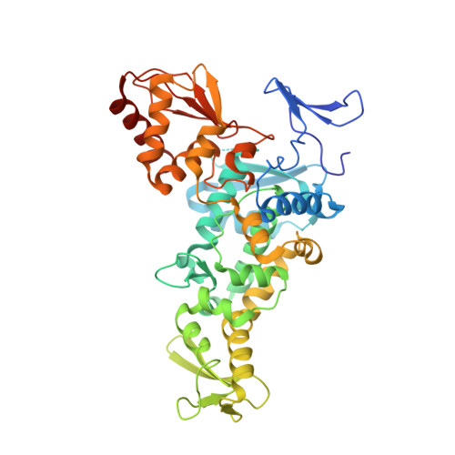
    Ubiquitination by HECT E3 enzymes regulates myriad processes, including tumor suppression, transcription, protein trafficking, and degradation. HECT E3s use a two-step mechanism to ligate ubiquitin to target proteins. The first step is guided by interactions between the catalytic HECT domain and the E2∼ubiquitin intermediate, which promote formation of a transient, thioester-bonded HECT∼ubiquitin intermediate. Here we report that the second step of ligation is mediated by a distinct catalytic architecture established by both the HECT E3 and its covalently linked ubiquitin. The structure of a chemically trapped proxy for an E3∼ubiquitin-substrate intermediate reveals three-way interactions between ubiquitin and the bilobal HECT domain orienting the E3∼ubiquitin thioester bond for ligation, and restricting the location of the substrate-binding domain to prioritize target lysines for ubiquitination. The data allow visualization of an E2-to-E3-to-substrate ubiquitin transfer cascade, and show how HECT-specific ubiquitin interactions driving multiple reactions are repurposed by a major E3 conformational change to promote ligation. DOI:http://dx.doi.org/10.7554/eLife.00828.001.


  • Organizational Affiliation
    • Department of Structural Biology , St Jude Children's Research Hospital , Memphis , United States.

Macromolecule Content 

  • Total Structure Weight: 125.23 kDa 
  • Atom Count: 7,944 
  • Modeled Residue Count: 1,004 
  • Deposited Residue Count: 1,078 
  • Unique protein chains: 3

Macromolecules

Find similar proteins by:|  3D Structure
Entity ID: 1
MoleculeChains  Sequence LengthOrganismDetailsImage
E3 ubiquitin-protein ligase RSP5
A, B
432Saccharomyces cerevisiae S288CMutation(s): 3 
Gene Names: MDP1NPI1RSP5SYGP-ORF41YER125W
EC: 6.3.2 (PDB Primary Data), 2.3.2.26 (UniProt)
UniProt
Find proteins for P39940 (Saccharomyces cerevisiae (strain ATCC 204508 / S288c))
Explore P39940 
Go to UniProtKB:  P39940
Entity Groups
Sequence Clusters30% Identity50% Identity70% Identity90% Identity95% Identity100% Identity
UniProt GroupP39940
Sequence Annotations
Expand
Reference Sequence
Find similar proteins by:  Sequence   |   3D Structure  
Entity ID: 2
MoleculeChains  Sequence LengthOrganismDetailsImage
Protein SNA3
C, D
24Saccharomyces cerevisiae S288CMutation(s): 1 
UniProt
Find proteins for P14359 (Saccharomyces cerevisiae (strain ATCC 204508 / S288c))
Explore P14359 
Go to UniProtKB:  P14359
Entity Groups
Sequence Clusters30% Identity50% Identity70% Identity90% Identity95% Identity100% Identity
UniProt GroupP14359
Sequence Annotations
Expand
Reference Sequence
Find similar proteins by:|  3D Structure
Entity ID: 3
MoleculeChains  Sequence LengthOrganismDetailsImage
Ubiquitin
E, F
83Homo sapiensMutation(s): 1 
Gene Names: UBCUBIQUITIN
UniProt & NIH Common Fund Data Resources
Find proteins for P0CG48 (Homo sapiens)
Explore P0CG48 
Go to UniProtKB:  P0CG48
PHAROS:  P0CG48
GTEx:  ENSG00000150991 
Entity Groups
Sequence Clusters30% Identity50% Identity70% Identity90% Identity95% Identity100% Identity
UniProt GroupP0CG48
Sequence Annotations
Expand
Reference Sequence

Experimental Data & Validation

Experimental Data

  • Method: X-RAY DIFFRACTION
  • Resolution: 3.10 Å
  • R-Value Free:  0.299 (Depositor), 0.297 (DCC) 
  • R-Value Work:  0.251 (Depositor), 0.252 (DCC) 
  • R-Value Observed: 0.253 (Depositor) 
Space Group: P 1 21 1
Unit Cell:
Length ( Å )Angle ( ˚ )
a = 83.046α = 90
b = 78.923β = 101.67
c = 96.722γ = 90
Software Package:
Software NamePurpose
ADSCdata collection
PHASERphasing
PHENIXrefinement
HKL-2000data reduction
HKL-2000data scaling

Structure Validation

View Full Validation Report


Entry History 

Deposition Data

Revision History  (Full details and data files)

  • Version 1.0: 2013-08-14
    Type: Initial release
  • Version 1.1: 2013-08-28
    Changes: Database references
  • Version 1.2: 2024-02-28
    Changes: Data collection, Database references