4LBL | pdb_00004lbl

Crystal structure of Human galectin-3 CRD K176L mutant in complex with a-GM3


Experimental Data Snapshot

  • Method: X-RAY DIFFRACTION
  • Resolution: 1.58 Å
  • R-Value Free: 
    0.190 (Depositor), 0.202 (DCC) 
  • R-Value Work: 
    0.153 (Depositor), 0.166 (DCC) 
  • R-Value Observed: 
    0.155 (Depositor) 

Starting Model: experimental
View more details

wwPDB Validation 3D Report Full Report

Validation slider image for 4LBL

This is version 2.1 of the entry. See complete history

Literature

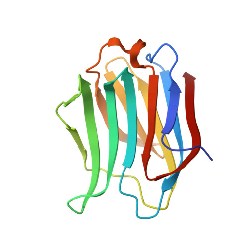
Galectin-3 interactions with glycosphingolipids.

Collins, P.M.Bum-Erdene, K.Yu, X.Blanchard, H.

(2014) J Mol Biology 426: 1439-1451

  • DOI: https://doi.org/10.1016/j.jmb.2013.12.004
  • Primary Citation Related Structures: 
    4LBJ, 4LBK, 4LBL, 4LBM, 4LBN, 4LBO

  • PubMed Abstract: 

    Galectins have essential roles in pathological states including cancer, inflammation, angiogenesis and microbial infections. Endogenous receptors include members of the lacto- and neolacto-series glycosphingolipids present on mammalian cells and contain the tetrasaccharides lacto-N-tetraose (LNT) and lacto-N-neotetraose (LNnT) that form their core structural components and also ganglio-series glycosphingolipids. We present crystallographic structures of the carbohydrate recognition domain of human galectin-3, both wild type and a mutant (K176L) that influenced ligand affinity, in complex with LNT, LNnT and acetamido ganglioside a-GM3 (α2,3-sialyllactose). Key structural features revealed include galectin-3's demonstration of a binding mode towards gangliosides distinct from that to the lacto/neolacto-glycosphingolipids, with its capacity for recognising the core β-galactoside region being challenged when the core oligosaccharide epitope of ganglio-series glycosphingolipids (GM3) is embedded within particular higher-molecular-weight glycans. The lacto- and neolacto- glycosphingolipids revealed different orientations of their terminal galactose in the galectin-3-bound LNT and LNnT structures that has significant ramifications for the capacity of galectin-3 to interact with higher-order lacto/neolacto-series glycosphingolipids such as ABH blood group antigens and the HNK-1 antigen that is common on leukocytes. LNnT also presents an important model for poly-N-acetyllactosamine-containing glycans and provides insight into galectin-3's accommodation of extended oligosaccharides such as the poly-N-acetyllactosamine-modified N- and O-glycans that, via galectin-3 interaction, facilitate progression of lung and bladder cancers, respectively. These findings provide the first atomic detail of galectin-3's interactions with the core structures of mammalian glycosphingolipids, providing information important in understanding the capacity of galectin-3 to engage with receptors identified as facilitators of major disease.


  • Organizational Affiliation
    • Institute for Glycomics, Griffith University Gold Coast Campus, Queensland 4222, Australia.

Macromolecule Content 

  • Total Structure Weight: 16.42 kDa 
  • Atom Count: 1,405 
  • Modeled Residue Count: 138 
  • Deposited Residue Count: 138 
  • Unique protein chains: 1

Macromolecules

Find similar proteins by:|  3D Structure
Entity ID: 1
MoleculeChains  Sequence LengthOrganismDetailsImage
Galectin-3138Homo sapiensMutation(s): 1 
Gene Names: LGALS3MAC2
UniProt & NIH Common Fund Data Resources
Find proteins for P17931 (Homo sapiens)
Explore P17931 
Go to UniProtKB:  P17931
PHAROS:  P17931
GTEx:  ENSG00000131981 
Entity Groups
Sequence Clusters30% Identity50% Identity70% Identity90% Identity95% Identity100% Identity
UniProt GroupP17931
Sequence Annotations
Expand
Reference Sequence

Oligosaccharides

Help  
Entity ID: 2
MoleculeChains Length2D Diagram GlycosylationD Interactions
N-acetyl-alpha-neuraminic acid-(2-3)-beta-D-galactopyranose-(1-4)-beta-D-glucopyranose
B
3N/A
Glycosylation Resources
GlyTouCan: G91237TK
GlyCosmos: G91237TK
GlyGen: G91237TK

Experimental Data & Validation

Experimental Data

  • Method: X-RAY DIFFRACTION
  • Resolution: 1.58 Å
  • R-Value Free:  0.190 (Depositor), 0.202 (DCC) 
  • R-Value Work:  0.153 (Depositor), 0.166 (DCC) 
  • R-Value Observed: 0.155 (Depositor) 
Space Group: P 21 21 21
Unit Cell:
Length ( Å )Angle ( ˚ )
a = 35.86α = 90
b = 57.61β = 90
c = 62.59γ = 90
Software Package:
Software NamePurpose
MOSFLMdata reduction
SCALAdata scaling
REFMACrefinement
PDB_EXTRACTdata extraction
Blu-Icedata collection
PHASERphasing

Structure Validation

View Full Validation Report


Entry History 

Deposition Data

Revision History  (Full details and data files)

  • Version 1.0: 2014-01-22
    Type: Initial release
  • Version 1.1: 2014-03-26
    Changes: Database references
  • Version 2.0: 2020-07-29
    Type: Remediation
    Reason: Carbohydrate remediation
    Changes: Atomic model, Data collection, Database references, Derived calculations, Structure summary
  • Version 2.1: 2023-09-20
    Changes: Data collection, Database references, Refinement description, Structure summary