4LAC

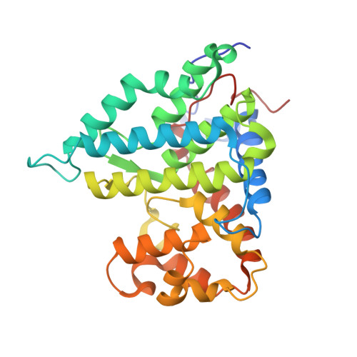
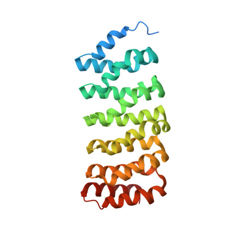
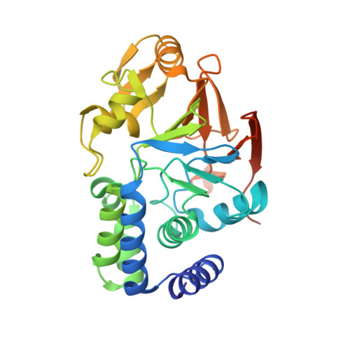
Crystal Structure of Protein Phosphatase 2A (PP2A) and PP2A phosphatase activator (PTPA) complex with ATPgammaS


Experimental Data Snapshot

  • Method: X-RAY DIFFRACTION
  • Resolution: 2.82 Å
  • R-Value Free: 0.242 
  • R-Value Work: 0.188 
  • R-Value Observed: 0.191 

Starting Models: experimental
View more details

wwPDB Validation   3D Report Full Report


Ligand Structure Quality Assessment 


This is version 1.3 of the entry. See complete history


Literature

Structural basis of PP2A activation by PTPA, an ATP-dependent activation chaperone.

Guo, F.Stanevich, V.Wlodarchak, N.Sengupta, R.Jiang, L.Satyshur, K.A.Xing, Y.

(2014) Cell Res 24: 190-203

  • DOI: https://doi.org/10.1038/cr.2013.138
  • Primary Citation of Related Structures:  
    4LAC

  • PubMed Abstract: 

    Proper activation of protein phosphatase 2A (PP2A) catalytic subunit is central for the complex PP2A regulation and is crucial for broad aspects of cellular function. The crystal structure of PP2A bound to PP2A phosphatase activator (PTPA) and ATPγS reveals that PTPA makes broad contacts with the structural elements surrounding the PP2A active site and the adenine moiety of ATP. PTPA-binding stabilizes the protein fold of apo-PP2A required for activation, and orients ATP phosphoryl groups to bind directly to the PP2A active site. This allows ATP to modulate the metal-binding preferences of the PP2A active site and utilize the PP2A active site for ATP hydrolysis. In vitro, ATP selectively and drastically enhances binding of endogenous catalytic metal ions, which requires ATP hydrolysis and is crucial for acquisition of pSer/Thr-specific phosphatase activity. Furthermore, both PP2A- and ATP-binding are required for PTPA function in cell proliferation and survival. Our results suggest novel mechanisms of PTPA in PP2A activation with structural economy and a unique ATP-binding pocket that could potentially serve as a specific therapeutic target.


  • Organizational Affiliation

    McArdle Laboratory, Department of Oncology, University of Wisconsin at Madison, School of Medicine and Public Health, Madison, WI 53706, USA.


Macromolecules
Find similar proteins by:  (by identity cutoff)  |  3D Structure
Entity ID: 1
MoleculeChains Sequence LengthOrganismDetailsImage
Serine/threonine-protein phosphatase 2A activatorA [auth B]308Homo sapiensMutation(s): 0 
Gene Names: PP2A Catalytic Subunit CPPP2R4PTPA
EC: 5.2.1.8
UniProt & NIH Common Fund Data Resources
Find proteins for Q15257 (Homo sapiens)
Explore Q15257 
Go to UniProtKB:  Q15257
PHAROS:  Q15257
GTEx:  ENSG00000119383 
Entity Groups  
Sequence Clusters30% Identity50% Identity70% Identity90% Identity95% Identity100% Identity
UniProt GroupQ15257
Sequence Annotations
Expand
  • Reference Sequence
Find similar proteins by:  (by identity cutoff)  |  3D Structure
Entity ID: 2
MoleculeChains Sequence LengthOrganismDetailsImage
PP2A Scaffold Subunit A, Truncated, an internal deletion of PP2A AB [auth A]258Homo sapiensMutation(s): 0 
Gene Names: PP2A Phosphatase Activator
UniProt & NIH Common Fund Data Resources
Find proteins for P30153 (Homo sapiens)
Explore P30153 
Go to UniProtKB:  P30153
PHAROS:  P30153
GTEx:  ENSG00000105568 
Entity Groups  
Sequence Clusters30% Identity50% Identity70% Identity90% Identity95% Identity100% Identity
UniProt GroupP30153
Sequence Annotations
Expand
  • Reference Sequence
Find similar proteins by:  (by identity cutoff)  |  3D Structure
Entity ID: 3
MoleculeChains Sequence LengthOrganismDetailsImage
Serine/threonine-protein phosphatase 2A catalytic subunit alpha isoform311Homo sapiensMutation(s): 0 
Gene Names: PP2A Scaffold Subunit APPP2CA
EC: 3.1.3.16
UniProt & NIH Common Fund Data Resources
Find proteins for P67775 (Homo sapiens)
Explore P67775 
Go to UniProtKB:  P67775
PHAROS:  P67775
GTEx:  ENSG00000113575 
Entity Groups  
Sequence Clusters30% Identity50% Identity70% Identity90% Identity95% Identity100% Identity
UniProt GroupP67775
Sequence Annotations
Expand
  • Reference Sequence
Experimental Data & Validation

Experimental Data

  • Method: X-RAY DIFFRACTION
  • Resolution: 2.82 Å
  • R-Value Free: 0.242 
  • R-Value Work: 0.188 
  • R-Value Observed: 0.191 
  • Space Group: P 21 21 21
Unit Cell:
Length ( Å )Angle ( ˚ )
a = 57.07α = 90
b = 100.186β = 90
c = 167.45γ = 90
Software Package:
Software NamePurpose
ADSCdata collection
PHASERphasing
REFMACrefinement
HKL-2000data reduction
HKL-2000data scaling

Structure Validation

View Full Validation Report


Ligand Structure Quality Assessment 


Entry History 

Deposition Data

Revision History  (Full details and data files)

  • Version 1.0: 2013-10-09
    Type: Initial release
  • Version 1.1: 2013-10-30
    Changes: Database references
  • Version 1.2: 2014-02-19
    Changes: Database references
  • Version 1.3: 2023-09-20
    Changes: Data collection, Database references, Derived calculations, Refinement description