4L9D

Crystal structure of the PKD1 domain from Vibrio cholerae metalloprotease PrtV


Experimental Data Snapshot

  • Method: X-RAY DIFFRACTION
  • Resolution: 1.10 Å
  • R-Value Free: 0.140 
  • R-Value Work: 0.109 
  • R-Value Observed: 0.111 

wwPDB Validation   3D Report Full Report


This is version 1.2 of the entry. See complete history


Literature

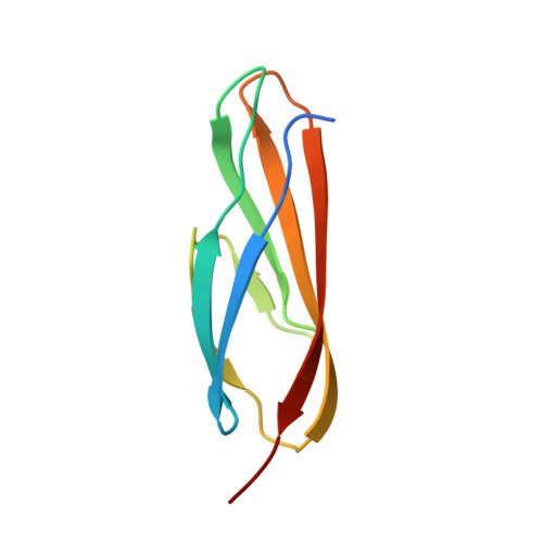
Calcium binding by the PKD1 domain regulates interdomain flexibility in Vibrio cholerae metalloprotease PrtV.

Edwin, A.Rompikuntal, P.Bjorn, E.Stier, G.Wai, S.N.Sauer-Eriksson, A.E.

(2013) FEBS Open Bio 3: 263-270

  • DOI: https://doi.org/10.1016/j.fob.2013.06.003
  • Primary Citation of Related Structures:  
    4L9D

  • PubMed Abstract: 

    Vibrio cholerae, the causative agent of cholera, releases several virulence factors including secreted proteases when it infects its host. These factors attack host cell proteins and break down tissue barriers and cellular matrix components such as collagen, laminin, fibronectin, keratin, elastin, and they induce necrotic tissue damage. The secreted protease PrtV constitutes one virulence factors of V. cholerae. It is a metalloprotease belonging to the M6 peptidase family. The protein is expressed as an inactive, multidomain, 102 kDa pre-pro-protein that undergoes several N- and C-terminal modifications after which it is secreted as an intermediate variant of 81 kDa. After secretion from the bacteria, additional proteolytic steps occur to produce the 55 kDa active M6 metalloprotease. The domain arrangement of PrtV is likely to play an important role in these maturation steps, which are known to be regulated by calcium. However, the molecular mechanism by which calcium controls proteolysis is unknown. In this study, we report the atomic resolution crystal structure of the PKD1 domain from V. cholera PrtV (residues 755-838) determined at 1.1 Å. The structure reveals a previously uncharacterized Ca(2+)-binding site located near linker regions between domains. Conformational changes in the Ca(2+)-free and Ca(2+)-bound forms suggest that Ca(2+)-binding at the PKD1 domain controls domain linker flexibility, and plays an important structural role, providing stability to the PrtV protein.


  • Organizational Affiliation

    Department of Chemistry, Umeå University, Umeå SE-901 87, Sweden ; Umeå Centre for Microbial Research (UCMR), Umeå University, Umeå SE-901 87, Sweden.


Macromolecules
Find similar proteins by:  (by identity cutoff)  |  3D Structure
Entity ID: 1
MoleculeChains Sequence LengthOrganismDetailsImage
Protease
A, B
88Vibrio cholerae M66-2Mutation(s): 0 
Gene Names: prtVVCM66_A0219
UniProt
Find proteins for C3LUP3 (Vibrio cholerae serotype O1 (strain M66-2))
Explore C3LUP3 
Go to UniProtKB:  C3LUP3
Entity Groups  
Sequence Clusters30% Identity50% Identity70% Identity90% Identity95% Identity100% Identity
UniProt GroupC3LUP3
Sequence Annotations
Expand
  • Reference Sequence
Small Molecules
Ligands 4 Unique
IDChains Name / Formula / InChI Key2D Diagram3D Interactions
PEG
Query on PEG

Download Ideal Coordinates CCD File 
J [auth B]DI(HYDROXYETHYL)ETHER
C4 H10 O3
MTHSVFCYNBDYFN-UHFFFAOYSA-N
CA
Query on CA

Download Ideal Coordinates CCD File 
F [auth B]CALCIUM ION
Ca
BHPQYMZQTOCNFJ-UHFFFAOYSA-N
CL
Query on CL

Download Ideal Coordinates CCD File 
D [auth A],
E [auth A],
I [auth B]
CHLORIDE ION
Cl
VEXZGXHMUGYJMC-UHFFFAOYSA-M
NA
Query on NA

Download Ideal Coordinates CCD File 
C [auth A],
G [auth B],
H [auth B]
SODIUM ION
Na
FKNQFGJONOIPTF-UHFFFAOYSA-N
Experimental Data & Validation

Experimental Data

  • Method: X-RAY DIFFRACTION
  • Resolution: 1.10 Å
  • R-Value Free: 0.140 
  • R-Value Work: 0.109 
  • R-Value Observed: 0.111 
  • Space Group: P 21 21 21
Unit Cell:
Length ( Å )Angle ( ˚ )
a = 41.8α = 90
b = 50.833β = 90
c = 67.297γ = 90
Software Package:
Software NamePurpose
XDSdata scaling
Auto-Rickshawphasing
REFMACrefinement
XDSdata reduction
SCALAdata scaling

Structure Validation

View Full Validation Report


Entry History 

Deposition Data

Revision History  (Full details and data files)

  • Version 1.0: 2013-07-10
    Type: Initial release
  • Version 1.1: 2013-08-14
    Changes: Database references
  • Version 1.2: 2024-02-28
    Changes: Data collection, Database references, Derived calculations